Musculoskeletal Imaging and Intervention

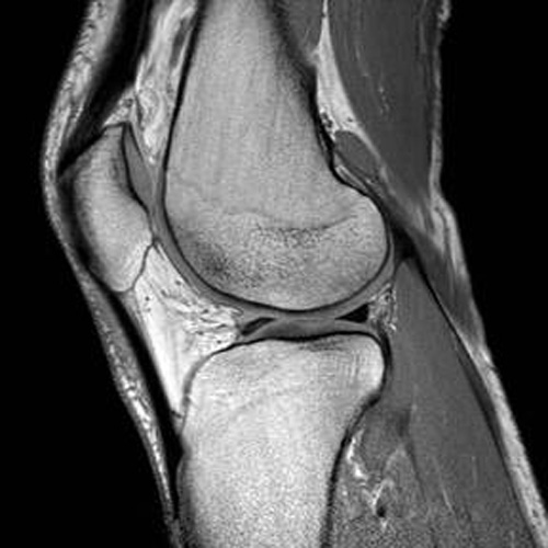
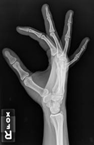
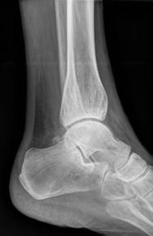
The musculoskeletal imaging section of OHSU Diagnostic Radiology provides comprehensive evaluation of bones, joints, muscles, and nerves. A complete spectrum of imaging modalities is employed, including MRI, CT, ultrasound, x-ray and fluoroscopy, and all studies are performed on state of the art equipment. We use the latest imaging techniques in order to address the specific concerns of our referring clinicians.
The section also maintains a robust musculoskeletal interventional practice, offering a wide array of services performed on a daily basis. These include fluoroscopic- and CT-guided joint injections; CT-guided spine, pelvic, and extremity bone and soft tissue biopsies; ultrasound-guided biopsies, aspirations, and injections; and CT-guided osteoid osteoma RFA (radiofrequency ablation).
All musculoskeletal radiologists are certified by the American Board of Radiology and have completed musculoskeletal fellowships. Meet the MSK Radiologist Team.
We work closely with referring clinicians in orthopedic surgery, sports medicine, physical medicine and rehabilitation, rheumatology, and other clinical services and in order to provide the best possible care for our patients. We hold weekly and monthly case conferences with referring clinicians to discuss patient care, imaging findings, and intervention.
In addition to our busy clinical service, the musculoskeletal faculty takes great pride in providing exceptional teaching in the radiology residency program and musculoskeletal radiology fellowship. Direct teaching occurs throughout the day at the workstation and in the procedure suite, complemented by weekly didactic and case conferences. A complete teaching curriculum ensures that our trainees are taught the most current topics and procedural techniques.
We also have several ongoing academic projects including scientific research, educational exhibits, international teaching efforts, and national, regional, and institutional lectures.
Fellowship
The Musculoskeletal (MSK) Radiology Fellowship is a non-ACGME program, requiring a 1 year commitment. Candidates must be Board Certified or Board Eligible radiologists and eligible for an unrestricted Oregon state medical license prior to beginning the fellowship. Candidates for fellowship are selected through the Musculoskeletal Fellowship Match. You can get additional information about the match and registration from the above link to the Society of Skeletal Radiology website.
The MSK section is composed of seven full-time faculty members and two fellows. Meet the team!
OHSU, Oregon’s only academic health center, is home to multiple state of the art CT and MRI scanners; the vast majority of joint imaging is performed on 3T scanners. The program has two fellowship positions and offers a comprehensive education in all facets of musculoskeletal radiology. The fellowship aims to produce well-rounded musculoskeletal radiologists in a collegial environment who are expert diagnosticians and interventionalists. These aims are achieved through a combination of teaching, clinical work and ample opportunities for research including protected academic/meeting time.
The fellows typically alternate in the diagnostics versus procedures roles every other week. The diagnostic MSK service interprets a large number of imaging studies daily, including radiography, MRI, CT, and ultrasound. The diverse case mix includes high-end sports MR and an active bone and soft tissue sarcoma service (Knight Cancer Institute). Fellows work side-by-side with attendings, reviewing cases throughout the day and receiving one-on-one teaching, gradually assuming more responsibilities as the year progresses. In addition to interpreting studies, fellows also develop a deep understanding of MRI and CT protocols by tailoring examinations for each patient, working closely with technologists to modify protocols according to specific clinical questions and troubleshooting challenging scenarios. While expected to be in the hospital while on daily clinical service, all fellows have home workstations allowing for increased flexibility including the ability to take call from home.
The section also performs a large number of procedures each week, including fluoroscopic-guided therapeutic injections/aspirations and MR arthrography, CT-guided biopsies/interventions including spine, miscellaneous injections, RF (radiofrequency) ablation, and ultrasound-guided biopsies/interventions. This includes all MSK-related procedures for pediatric patients via Doernbecher Children’s Hospital. Fellows are an integral part of this service and work closely with referring clinicians to decide on appropriate plans of action and patient management.
The Musculoskeletal section is actively engaged with referring clinical colleagues and our fellows are often the first line of communication between services. These critical relationships are maintained and strengthened through weekly Rheumatology-Radiology and Orthopedic Oncology-Pathology-Radiology conferences and conferences which provide valuable insight into the needs of referring providers as well as feedback on imaging interpretation and procedures. In addition to daily clinical teaching and multidisciplinary conferences, the MSK fellowship curriculum includes a monthly lecture series dedicated to covering highly subspecialized topics to ensure our fellows have as complete an education as possible. Finally, the fellowship training is supplemented with journal club, arthroscopy correlation, biopsy rad-path correlation and hands-on ultrasound didactics.
Fellows leave the program well-prepared for both private practice and academic settings with strong procedural skills, a balanced fund of knowledge and a solid framework for approaching even the most difficult problems in musculoskeletal imaging.
A message from the program director

Welcome to PDX, Portlandia, Stumptown, the City of Roses, or my personal favorite, Rip City! No matter the name, the Pacific Northwest or as loving called by the locals, Pacific Wonderland, is an incredible place to call home whether for a year or a career.
Thank you for your interest in OHSU's Musculoskeletal Imaging and Intervention Fellowship. Our goal is to help fellows achieve their full potential and graduate the program well prepared for both private practice and academic settings with strong procedural skills, a well-rounded fund of knowledge, and a solid framework for approaching even the most difficult problems in musculoskeletal imaging.
This learning occurs in a collegial environment among seven full-time faculty members who are passionate educators and dedicated clinicians. Our fellows are critical components to our musculoskeletal team and encouraged from day one to take ownership of their education with graduated responsibilities escalating throughout the fellowship year.
If I had to speak to one strength of our fellowship, I would highlight balance. By balance, I mean not just from a solid mix of clinical knowledge and procedural skills, but also from the all important work-life perspective. Our fellows have ample academic and elective time as well as the vast majority of their weekends free of clinical duties. This allows for personal growth both in and outside of the reading room as the journey of learning is a lifelong endeavor. This journey is a finite one, why not spend a portion of yours in the Pacific Wonderland?
Barry Hansford, M.D.
Annual total imaging volumes
Radiography – 57000 I MSK CT – 1500 I MSK MRI – 3700 I MSK Diagnostic US – 75 I Osteoid Osteoma RFA – 8 I CT-guided bone/soft tissue biopsy – 150 I US-guided biopsy – 50 I Fluoroscopic-guided injections/aspirations – 500
- Fellowship Lecture Series- Two to three Thursdays per month at noon. MSK faculty provide 30-45 minute lectures based on the musculoskeletal radiology fellowship curriculum.
- Musculoskeletal Case of the Day- Daily 4pm interesting case conference for residents and fellows.
- Resident Lecture Series- Approximately 30 resident lectures are given by MSK faculty to residents and fellows. Near the end of fellowship, fellows will usually give at least one lecture to the residents with faculty support.
- Journal Club- Approximately 4-6 meetings per year discussing pertinent/hot topics in the MSK literature.
- Hands-On MSK Ultrasound Didactics- Approximately 4-6 faculty-guided joint focused US didactics per year.
- Radiology-Pathology Correlation- Quarterly conference reviewing all MSK bone and soft tissue biopsies for concordance, QA, and interesting cases/techniques.
- Western MSK Fellowship Cross-Institutional Collaboration- Rotating 12-16 lectures per year on select Friday mornings given by OHSU, University of Utah, University of Colorado, and University of Washington faculty
Fellows are assigned to cover tumor board and multiple working conferences with faculty throughout the year:
- Sarcoma Tumor Board- Wednesdays at 12pm
- Rheumatology Interesting Cases- Wednesdays at 10:15am
- Orthopedic Radiology-Arthroscopy- Bi-annual at 6:30am
- MSK Trauma-Radiology- Bi-annual at 12:00pm
- General Trauma-Radiology- 4-6 times per year on Wednesdays at 7:30am
- Fellows receive 26 days of PTO each year and one Juneteenth day, from which both sick and vacation time is used. An additional Extended Illness bank is also established for circumstances when the PTO bank is exhausted.
- MSK fellow salary is commensurate with PGY Year and based on the AAMC Western State Mean Trainee Figures, updated each January
- Each fellow earns 10 days of CME conference time, more meeting time may be approved at the department’s discretion if a fellow is presenting at a meeting.
- Fellows are currently offered funds, up to $3000/year, to reimburse for educational materials, books, and CME or board review courses.
- Fellows take approximately 6-8 general call weekends a year starting in September for which they cover all STAT and inpatient radiographs. Fellows also take approximately 8-10 short call shifts per year from 5pm-9pm in which they cover non-neuroradiology ED plain films and cross-sectional imaging (CT and US). Finally, fellows take part in the MSK backup spine biopsy call pool approximately 6-8 weekends a year which runs concordantly with the assigned general call weekends.
- Opportunities for fellow moonlighting are available via the VA Healthcare System
Half-days per week starting in September which can be combined for one full academic day every two weeks. More academic is available at faculty discretion given fellow interest/involvement in scholarly projects.
Fellows are encouraged, but not required to engage in a scholarly project. Ample opportunity exists to work on abstracts for presentation at national and international meetings or manuscripts for publication.
Each fellow is given two weeks of protected time to rotate through other radiology sections or departments during the second half of the academic year.
- Primary lymphoma of bone of the little finger: a case report and review of the literature. Skeletal Radiol. 2024 Jan 16. doi: 10.1007/s00256-024-04576-9. Epub ahead of print. PMID: 38225403
- Cubonavicular coalition: magnetic resonance imaging findings and associated pathology of a rarely reported condition in 27 patients. Acad Radiol. 2023 Sep 6:S1076-6332(23)00410-5. doi: 10.1016/j.acra.2023.08.011. Epub ahead of print. PMID: 37684180
- Beyond schwannomas and neurofibromas: a radiological and histopathological review of lesser-known benign lesions that arise in association with peripheral nerves. Skeletal Radiol. 2022 Oct 25. doi: 10.1007/200256-022-04207-1. Epub ahead of print. PMID: 36280619
- Fluoroscopic-guided procedures of the lower extremity. Skeletal Radiol. 2022 Aug 5. doi: 10.1007/s00256-022-04139-w. Epub ahead of print. PMID: 35930079
- Unusual case of Anti-SRP necrotizing myopathy with predominant distal leg weakness and atrophy. Neuromuscul Disord. 2021 Nov 19:S0960-8966(21)00709-4. doi: 10.1016/j.nmd.2021.11.010. Epub ahead of print. PMID: 35031192
- Concepts in musculoskeletal bone and soft tissue biopsy. Semin Musculoskeletal Radiol. 2021, Epub 2021. DOI: 10.1055/s-0041-1735471. Epub 2021 Dec 22. PMID: 34937112
- Tenosynovial giant cell tumor-diffuse type, treated with a novel colony-stimulating factor inhibitor, pexidartinib: initial experience with MRI findings in three patients. Skeletal Radiol. 2021. Sep 29. doi: 10.1007/s00256-021-03924-3. Online ahead of print. PMID: 34586485
- Novel EGFL7-FOSB fusion in pseudomyogenic hemangioendothelioma with widely metastatic disease. Histopathology 2021; In production. DOI: 10.1111/his.14349. PMID:32815039
- Multimodality pitfalls of wrist imaging with a focus on magnetic resonance imaging: what the radiologist needs to know. Top Magn Reson Imaging 2020; 29:263-272. DOI: 10.1097/RMR.0000000000000254. PMID:33021577
- Percutaneous image-guided sternal biopsy: a cross-institutional retrospective review of diagnostic yield and safety in 50 cases. Skeletal Radiol. 2021;50:495-504. DOI: 10.1007/s00256-020-03587-6. PMID: 32815039
- Untreated plasmacytoma of bone containing macroscopic intralesional fat and mimicking intraosseous lipoma: a case report and review of the literature. Clin Imaging. 2020;64:18-23. DOI:10.1017/j.clinimag.2020.03.003. PMID: 32208179
- Avulsion injuries and tendon ruptures of the hand and wrist: A review of frequently encountered and uncommon radiologic diagnoses. RadioGraphics. 2020;40:163-180. DOI: 10.1148/rg.2020190085. PMID: 31917655
- Pearls and pitfalls for soft-tissue and bone biopsies: cross institutional review. RadioGraphics. 2020;40:266-290. DOI 10.1148/rg.2020190089. PMID: 31917660
- SSR white paper on- optimal approaches to imaging inflammation associated arthritis. Society of Skeletal Radiology, Santa Fe NM, March 30-April 2, 2025
- MRI of Morton’s neuroma post-cryoablation: a previously undescribed distinctive MR imaging appearance at the distal metatarsals in three patients. Society of Skeletal Radiology, Santa Fe NM, March 30-April 2, 2025
- Post-traumatic entities mimicking bone and soft tissue neoplasms: what the radiologist needs to know. Radiological Society of North America, Chicago IL, Dec. 1-5, 2024
- Risk versus benefit of percutaneous image-guided biopsy for peripheral nerve sheath tumors: updated recommendations for who, what, when, and how. Radiological Society North America, Chicago IL, Dec. 1-5, 2024
- Diagnostic yield of extraosseous soft tissue component in evaluation of bony lesions with extraosseous soft tissue extension. Society of Interventional Oncology, Long Beach CA, Jan. 25-29, 2024
- Thumb distal phalangeal pseudolesion: imaging findings and association of an underappreciated normal variant that may mimic osteolytic pathology. Society of Skeletal Radiology, San Juan PR, April 4-7, 2024 (Certificate of merit award winner)
- Intraosseous lesions with extraosseous extension of tumor: Does biopsying only the soft tissue component result in an acceptable diagnostic yield? Society of Skeletal Radiology, San Juan PR, April 4-7, 2024
- Thumb distal phalangeal pseudolesion: imaging findings and associations of an underappreciated normal variant that may mimic osteolytic pathology. Radiological Society North America, Chicago IL, Nov. 26-Nov 30, 2023
- Pediatric low-grade osteosarcomatous dedifferentiation of an atypical lipomatous tumor: case of the day award winner/presentation. Society of Skeletal Radiology, Savannah GA, March 12-15, 2023
- Novel trans-sacral approach for CT-guided percutaneous needle biopsy in patients with suspected L5-S1 discitis osteomyelitis. Society of Skeletal Radiology, Savannah GA, March 12-15, 2023
- Pseudomyogenic hemangioendothelioma: case of the day award winner/presentation. Society of Skeletal Radiology, Coronado CA, March 13-16, 2022
- Beyond schwannomas and neurofibromas: a review of benign and malignant lesions that arise in association with nerves. Society of Skeletal Radiology, Coronado CA, March 13-16, 2022 (Certificate of merit award winner)
- MRI of the popliteus tendon: age-related changes in size and signal in asymptomatic patients and association with osteoarthrosis. Society of Skeletal Radiology, Coronado CA, March 13-16, 2022
- Current musculoskeletal radiology education: survey of residency program directors in the United States. Society of Skeletal Radiology, Coronado CA, March 13-16, 2022
- Beyond schwannomas and neurofibromas: a review of benign and malignant lesions that arise in association with nerves. Radiological Society North America, Chicago IL, Nov. 28-Dec 2, 2021 (Certificate of merit award winner)
From the slopes of Mt. Hood to the shores of Cannon Beach, Oregon is referred to as a “Pacific Wonderland” for good reason and Portland is perfectly situated to take full advantage of all it. Here is just a small sampling of the innumerable things us Portlanders love about where we live, work, and play. Learn more!
- John Ward, MD, 2024. Body Fellowship University of Washington
- Veronica Krull, MD, 2024. Memorial Hermann Imaging Center, Sugar Land, TX
- Dallas Blanco, DO 2023. Acadiana Radiology Group, APMC, Lafayette, LA
- Ahmed Gabr, MD 2022. Assistant Professor of Radiology OHSU
- Alexei Ku, MD, 2021. Kaiser Permanente, Portland, OR
- Brendan Case, MD, 2021. Kaiser Permanente, Portland, OR
- David Feigal, MD, 2020. Carilion Clinic, Roanoke, VA
- Rex Holliday, MD, 2020. Private Practice, Gillette, WY
- Jaryd Yee, MD, 2019. Renaissance Imaging Medical Associates INC, Maui, HI
- Kevin He, MD, 2019. Mercy One, Waterloo, IA
Please inquire with Melissa Reed, Administrative Coordinator, at reedmeli@ohsu.edu if interested.
Application information
Our program is filled through June 2027. We will begin accepting applications for 2 positions for our 2027/28 Fellowship Year beginning November 1, 2025. Interviews will take place in early 2026. Our program will participate in the NRMP to match our fellowship positions. Learn more about the fellowship match on the Society of Skeletal Radiology Website.
Required application materials are:
- One completed and signed universal application form
- Curriculum vitae
- Personal statement
- Three letters of recommendation*
- Photocopy of the Dean's letter from your medical school or ECFMG certificate
- USMLE Step 1, 2, 3 scores
*If emailed, letters of recommendation must be sent directly from the recommender (or assigned admin) to Melissa Reed. If a hard copy is mailed, it must be in a sealed envelope.
Send your application packet by email to Melissa Reed.
Address the packet to:
Barry Hansford, M.D. - MSK Radiology Fellowship Director
ATTN: Melissa Reed
Mail Code: L-340
Oregon Health & Science University
3181 S.W. Sam Jackson Park Road
Portland, OR 97239
A confirmation letter or email will be sent to you once all documents are received.
For additional information or questions, contact Melissa Reed.
Gender Equity in Academic Radiology
OHSU School of Medicine Diversity and Equity
OHSU has paused recruitment for applicants who would require a new H-1B visa, in response to recent federal changes. If you have an H1-B visa currently or do not need a visa (e.g. are a green card holder), we are still accepting applications.
Routine and advanced imaging provided

Fracture Imaging | Ligament Imaging | Muscle and Tendon Imaging | Joint Instability Evaluation | Cartilage Evaluation | Rheumatologic Disease Imaging | Spine Alignment Evaluation | Bone and Soft Tissue Tumor Imaging | MR and CT Arthrography | Diagnostic and Therapeutic Joint Injections | Hardware Assessment | Bone Biopsy | Spine Biopsy | Muscle and Other Soft Tissue Biopsy | Osteoid Osteoma RF (Radiofrequency) Ablation | Iliopsoas Bursography | Muscle and Tendon Ultrasound | Imaging of Peripheral Nerves
