Ultrasound and Vascular Ultrasound
Ultrasounds use sound waves to create images of the inside of the body. Ultrasounds, also called sonograms, are painless. They don’t use radiation.
Doctors use ultrasounds to:
- Help diagnose conditions
- Help guide procedures
- Check how a baby is growing
Doctors use vascular ultrasounds to check how blood is flowing.
We have expertise in:
- Several types of prenatal ultrasound, including ultrasound for high-risk pregnancy
- Contrast-enhanced ultrasound for more detailed images
- Ultrasounds for breast conditions
- Ultrasounds for patients who have had or are about to have an organ transplant
- Liver elastography, to check liver health
- Transcranial doppler, to check blood flow in the brain
How ultrasound works
Before your appointment
You’ll get instructions through MyChart. Please follow them, or we may have to reschedule your appointment.
At your appointment
You may be asked to remove items, including jewelry, that could disrupt the scan. If you need to remove clothing, we’ll give you a gown.
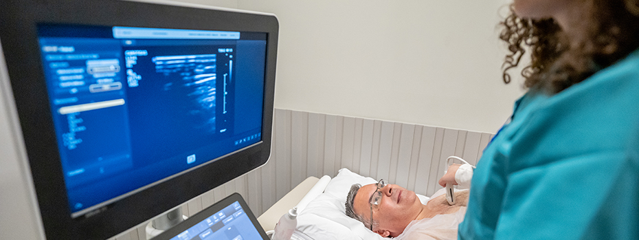
An ultrasound technician will place gel on a device called a transducer. The tech will place the transducer on your skin and move it around to get multiple images. The gel is harmless and washes off easily.
After your appointment
You’ll get results through MyChart.
Locations
OHSU Diagnostic Imaging Clinic, Marquam Hill (OHSU Hospital)
OHSU Hospital, 10th floor
3181 S.W. Sam Jackson Park Road
Portland, OR 97239
OHSU Diagnostic Imaging Clinic, Marquam Hill (Physicians Pavilion)
Physicians Pavilion, fourth floor
3270 S.W. Pavilion Loop
Portland, OR 97239
OHSU Diagnostic Imaging Clinic, South Waterfront
Center for Health & Healing, Building 1, third floor
3303 S. Bond Ave.
Portland, OR 97239
OHSU Diagnostic Imaging Clinic, Beaverton
15700 S.W. Greystone Court
Beaverton, OR 97006
Free parking for patients and visitors
Refer a patient
- Refer your patient to OHSU.
- Order diagnostic imaging.
- Call 503-494-4567 to seek provider-to-provider advice.
- Share your imaging.