Cardiothoracic Imaging
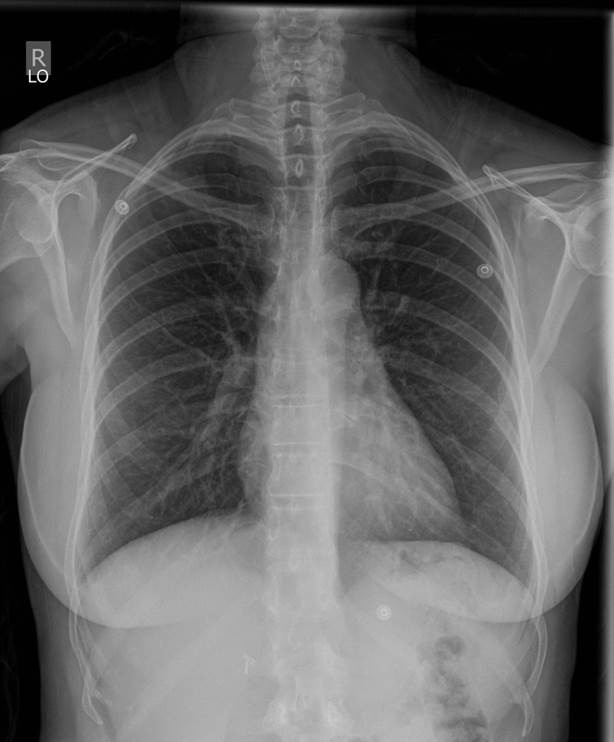
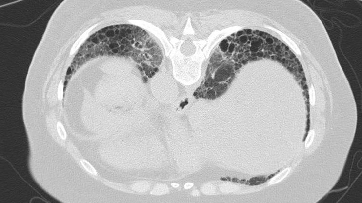
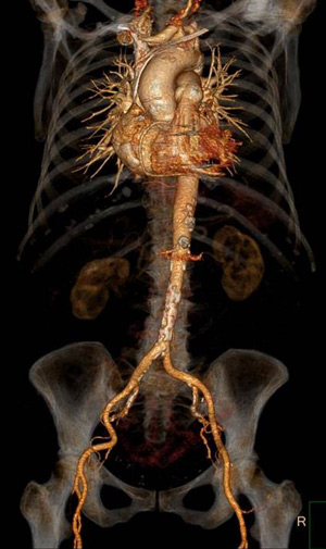
Cardiothoracic Imaging at OHSU offers basic to advanced imaging care. We use standard radiographs and computed tomography (CT) on a daily basis. Magnetic Resonance Imaging (MRI) is occasionally used for more advanced problem solving and mediastinal diseases or in younger patients with radiation exposure issues.
The chest radiograph remains the mainstay of our diagnostic imaging division, sufficient to evaluate most thoracic problems. Our CT scans are used to assess for the more persistent or serious problems that are not adequately evaluated on the standard radiographs. We also commonly use thin section scanning for fine detailed examination of the lungs and pulmonary vessels. Meet the Cardiothoracic Radiologist Team.

Fellowship
As the only academic health center in in the state of Oregon, Oregon Health & Science University provides a full spectrum of care to a diverse and growing population. OHSU is home to numerous research centers and institutes, including one of the first Clinical and Translational Science Award-funded programs in the country (Oregon Clinical and Translational Research Institute), the Advanced Imaging Research Center, the Dotter Interventional Institute and both the Knight Cancerand Knight Cardiovascular Institute. The Knight Cancer Institute is the only NCI-designated Cancer Center in Oregon.
The OHSU cardiothoracic imaging section is staffed by fellowship-trained cardiothoracic radiologists who cover all aspects of cardiac and thoracic imaging including radiography, CT/CTA, MRI/MRA and thoracic biopsy procedures. There is plenty of opportunity for institutional interdisciplinary interactions through multidisciplinary conferences and tumor boards, including family medicine, pulmonology and critical care, pathology, thoracic and cardiac surgery, medical oncology and radiation medicine. We encourage fellows to participate in collaborative research. Collaborations with the Knight Cancer and Knight Cardiovascular Institutes allow for cutting edge technologies and research, both clinical and preclinical. We also encourage and offer support for fellows to present at conferences through protected academic time and a discretionary fund. Our department's advanced facilities and expert mentors will give fellows the skills required to become first-rate cardiothoracic radiologists.
Application information
The Cardiothoracic Fellowship Program will be recruiting two fellows for the July 2027-June 2028 academic year. Applications will be accepted starting November 1, 2025.
To apply, please email the following documents to Audrey Walsh:
- Curriculum Vitae (CV)
- Personal Statement
- Three letters of recommendation
- MSPE (Medical Student Performance Evaluation), or transcripts from your medical school
- Official Transcript of USMLE Steps 1, 2, and 3
- ECFMG certificate (only applies to International Medical Graduates)
If you have questions about our program, please email Administrative Coordinator, Audrey Walsh and Cardiothoracic Fellowship Program Director, Dr. En-Haw Wu.
OHSU has paused recruitment for applicants who would require a new H-1B visa, in response to recent federal changes. If you have an H1-B visa currently or do not need a visa (e.g. are a green card holder), we are still accepting applications.
Routine and advanced imaging services provided
Adult congenital heart disease | Congenital heart disease | Coronary artery anomalies | Cardiac tumors | Cardiomyopathy | Acute and chronic myocardial infarction | Cardiac tumors | Cardiomyopathy | Coronary artery calcium scoring | Coronary artery disease | Coronary artery bypass graft patency | Heart failure | Myocardial disease; infiltrative diseases, iron imaging | Myocardial viability | Myocardial perfusion | Adenosine stress myocardial perfusion | Pericardial disease | Pulmonary vein imaging | Valve disease
Outpatient imaging for acute or chronic diseases | Diffuse lung diseases | Airway diseases: Bronchiectasis and Small airway problems | Pulmonary vascular diseases: Acute and Chronic | Pleural diseases | Lung malignancies | Lung Cancer Screening | Pulmonary infections | Mediastinal diseases | Aortic diseases |Lung biopsies for cancer or infection | Pleural effusion sampling | Multidisciplinary evaluation of lung cancer | Lung and Bone marrow transplant evaluation | Unusual/Rare thoracic diseases | Diaphragmatic diseases | Pulmonary Hypertension evaluation


The Cardiac Imaging Section of Diagnostic Radiology is a multidisciplinary team of Radiologists and Cardiologists. We perform detailed anatomic, functional, and physiologic imaging of the coronary arteries, myocardium, cardiac chambers, valves, aorta, pulmonary arteries, and pericardium using cardiac magnetic resonance imaging (MRI) and computed tomography (CT) in adults and pediatric patients suffering from a broad range of congenital and acquired cardiac diseases.
All radiologists are certified by the American Board of Radiology; in addition, they have completed subspecialty training in the area of both thoracic and cardiac imaging. Cardiothoracic Imaging works closely with many clinical departments, such as the Knight Cardiovascular Institute, Pulmonary Medicine and the Knight Lung Cancer team. We participate in multiple daily & weekly multidisciplinary conferences and rounds with these clinical colleagues, offering a unique approach to patient care. Faculty presents at numerous local, state and national conferences as well as invited national and international lectures. Our division works closely with the OHSU School of Medicine in regards to teaching and research.