Neuroradiology
The Neuroradiology Section of Diagnostic Radiology offers comprehensive CT and MRI services for imaging of the brain, spine, and head & neck in both adult and pediatric patients. Our advanced imaging capabilities paired with subspecialty-trained neuroradiologists provides accurate diagnosis of a range of diseases including stroke, tumor, trauma, demyelinating disease, epilepsy, headache, neurodegenerative disease, degenerative spine disease, and other complex neurologic conditions. Additionally, our faculty perform image-guided spine procedures including diagnostic and therapeutic lumbar punctures, and myelography. Our fleet of 12 MRI scanners and 8 CT scanners are distributed among several locations in the Portland metro area, allowing for convenient scheduling for our patients.
Neuroradiology plays a vital role in the multidisciplinary care model at OHSU. We collaborate closely with clinical partners across neurology, neurosurgery, interventional neuroradiology, ENT surgery, orthopedic surgery, vascular surgery, internal medicine, and other specialties. Each examination is carefully tailored to the specific diagnostic questions posed by the referring clinicians. The Neuroradiology service is located on the 10th floor of the main hospital.
All healthcare providers are certified by the American Board of Radiology. Our Neuroradiologists have completed subspecialty fellowship training, and have acquired additional CAQ board certification in Neuroradiology. Meet the Neuroradiologist Team.
Fellowship
The Diagnostic Neuroradiology Fellowship is an ACGME accredited program, requiring a one- or two-year commitment. Candidates must be Board Certified or Board Eligible radiologists. Selection occurs through the National Residency Matching Program (NRMP). You can get additional information about the match and registration from the NRMP website.
The goals of this fellowship are two-fold: to train neuroradiologists with the highest level of clinical expertise, and to cultivate future leaders in academic neuroradiology. To achieve these goals, fellows engage in comprehensive clinical, research, and educational activities through their training.
Our fellows gain experience across all imaging modalities used to evaluate neurologic disease. During the fellowship year, our fellows interpret a high volume of CT and MRI studies, perform lumbar punctures and myelography, and have exposure to ultrasound, nuclear medicine and radiographic studies. Fellows will receive training in and interpret advanced imaging techniques at OHSU including CT and MRI perfusion, functional MRI, and PET/MRI.
Our program is a well-balanced academic training program that encompasses all aspects of both adult and pediatric neuroradiology. As the only academic center in Oregon, OHSU serves a large and diverse catchment area, offering exposure to a broad array of both foundational and highly complex clinical cases. OHSU provides a rich multidisciplinary learning environment for neuroradiology fellows, in collaboration with OHSU Neurology and other subspecialty departments, including centers specializing in stroke, epilepsy, and Alzheimer’s disease. The fellowship includes focused time in three key subspecialties of neuroradiology: Neurosurgery, Head and Neck Imaging, and Pediatric Neuroradiology.
Interventional neuroradiologic procedures are also performed at state-of-the-art levels with the Dotter Institute at OHSU, and neuroradiology fellows will actively participate in these diagnostic angiographic procedures with the neurointerventional team. Additional partnership with neuroscience researchers, the Oregon Regional Primate Center, leading MR physicists and image analysis experts, offer a unique opportunity for advanced academic training.
Connect with us on social media
A message from the program director
I warmly welcome you to the enriching OHSU Neuroradiology Fellowship! OHSU is located in the vibrant city of Portland, Oregon, and surrounded by the beauty of the Pacific Northwest. Our close-knit community offers a strong foundation in adult and pediatric neuroradiology, trauma, head & neck imaging, spine, and procedural skills. With supportive co-fellows and faculty, the program is tailored to your individual needs and offers an ideal work-life balance through flexible schedules and diverse shift options. Our dynamic curriculum, featuring weekly fellow-dedicated lectures and hands-on experience with advanced imaging techniques, will sharpen your diagnostic skills and foster lasting professional relationships. I am proud to highlight our state-of-the-art tools, and I am excited to share our nurturing environment that supports your transition to independent practice and a rewarding career in neuroradiology. I look forward to welcoming you into our OHSU family and joining you on this incredible journey.
Best,
Jaclyn Thiessen, M.D.

Approximate annual total imaging volumes
Neuro CT - 27,000 | Neuro MRI - 21,200 | Lumbar Punctures/ Myelograms - 600
Lectures and conferences
- Anatomy Lecture Series – Weekly 30 min fast paced lectures reviewing core neuroanatomy within the first few months of fellowship.
- Fellowship Lecture Series –Weekly 30 min didactic lectures of advanced topics, following the neuroradiology fellowship curriculum.
- Interesting Case series – Weekly 30 min review of notable cases from the prior week and faculty teaching files.
- Resident Lecture Series – Approximately 30 one-hour resident lectures delivered by neuroradiology faculty to residents and fellows (Near the end of the fellowship, fellows typically give at least one lecture to the residents with faculty guidance).
Neurointerventional rotation
Fellows spend two weeks on the neurointerventional rotation performing diagnostic and therapeutic procedures. Fellows may spend additional time on rotation if desired or if they have a more focused interest in neurointerventional procedures.
Call responsibilities
Fellows take call with faculty for one week at a time beginning on Friday, approximately every four weeks. Call frequency remains every four weeks, even if there are 3 fellows. Fellows do not take neurointerventional call.
Tumor boards and working conferences
Fellows are assigned to cover various Multidisciplinary tumor boards and Case conferences, with faculty oversight, throughout the year. These high-yield conferences enhance contextual learning, and promote the development of sustained collaborative relationships with our referring clinicians. These include:
- Neurology/Neurosurgery
- Adult Brain Tumor Board
- Spine tumor board
- Neuro ICU Case Conference
- Adult Epilepsy Conference
- Pediatric Neuroradiology
- Pediatric Brain Tumor Board
- Pediatric Neurology Interesting Case conference
- Pediatric stroke case conference
- Pediatric Epilepsy Conference
- Head and Neck
- ENT Tumor Board
- Neuro-ophthalmology interesting case conference
- Skull base tumor board
- Neuro Vascular malformations
Projects
Fellows are expected to participate in at least one scholarly activity, and one Quality Improvement (PQI) activity per year.
Application information
Our fellowship program is full through 2026-2027. We will be accepting applications for 2027-2028 through ERAS.
To apply, please upload to ERAS:
- Curriculum vitae
- Personal statement
- Three letters of recommendation
- Photocopy of the Dean's letter from your medical school or ECFMG certificate
- USMLE Step 1, 2, 3 board scores
For additional information or questions, contact Kim Meeder, Neuroradiology Fellowship Coordinator.
OHSU has paused recruitment for applicants who would require a new H-1B visa, in response to recent federal changes. If you have an H1-B visa currently or do not need a visa (e.g. are a green card holder), we are still accepting applications.
For More Information on OHSU Trainee Policies. Please visit our GME page.
This is information for our diagnostic neuroradiology fellowship. If you are interested in a neurointerventional fellowship please contact Ryan Priest, Fellowship Director or visit the Charles T. Dotter Department of Interventional Radiology Page.
Routine and advanced imaging provided
Brain Tumor Evaluation | Aneurysm Evaluation | Seizure Evaluation | Traumatic Brain Injury | Spinal Injury | Head and Neck Imaging | Dynamic Pituitary Imaging | Spine Imaging | Pediatric Brain and Spine Imaging | Congenital Malformations | Lumbar punctures | Cervical, Thoracic, and Lumbar Myleography | Magnetic Resonance Angiography (MRA) | Magnetic Resonance Venography (MRV)
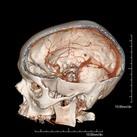
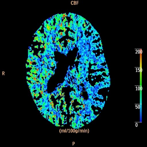
How it works: CT Perfusion evaluates how well blood is flowing to the brain. A small bolus of iodine based intravenous contrast is injected rapidly through a vein, and then multiple low dose CT scans are taken through the same area of the brain to evaluate how the contrast bolus flows through a portion of the brain over time. The data is then processed with advanced perfusion software.
Equipment: Perfusion studies are usually performed on a Philips 64 or 256 channel CT scanner. OHSU is an ACR accredited CT facility.
Benefits: CT Perfusion is a fast, minimally invasive method of evaluating cerebral perfusion. Reasons for the exam include evaluation of brain perfusion after stroke, temporary ischemic attack (TIA), and/or occlusion or narrowing of a major intracranial vessel.
Exam Preparation: Preparation for a CT perfusion study is the same as for a CT angiogram study. The technologist will interview you for contraindications to contrast such as allergy or kidney problems. If you have had an allergy to iodine based contrast in the past, you should discuss this with your physician before the study. In some situations, an alternative imaging method may be considered, or you may receive medication that needs to be taken before the study to reduce your risk of reaction. In some situations, kidney function labs may also be checked before your study. An IV will be placed, usually in the arm.
What to expect: Once you are positioned on the table and the contrast injector tubing is attached to your IV, a localizer scan will be taken, which takes a few seconds. The actual perfusion scan then takes 50 seconds. It is very important that your head remains still during the scan time. The technologist will check the images before you leave the room. In some situations, a non contrast CT or CTA study will be performed first. If a CTA is performed first, you will have to wait about 10 minutes between CTA and perfusion scans, to let the first bolus of contrast dissipate. After you have left, the perfusion data will be processed and analyzed, and a board certified neuroradiologist will review the study.
Adverse reactions to contrast materials are uncommon, but can range from mild to severe. Severe reactions are very uncommon. Further information about the risks and benefits of x-rays and contrast material can be found here.
Recent FDA publications have warned of the risks associated with high x-ray doses administered during CT perfusion exams at certain facilities. OHSU uses the latest technology to reduce the x-ray exposure as much as possible while maintaining a high quality examination.
Content by Dr. Louis P. Riccelli
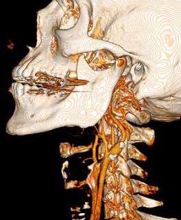
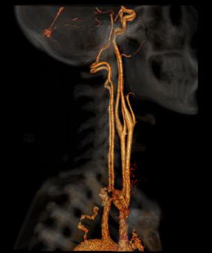
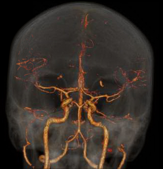
How it works: CT angiography (CTA) evaluates the major vessels of the head, neck or both. An iodine based contrast agent is rapidly injected through an IV placed in a vein, usually in the arm. A CT scan uses x-rays to acquired images as the contrast bolus passes through the arteries. The data can then be reviewed in multiple planes, and 3 dimensional images can also be created for review.
Equipment: Most commonly, a 64 channel Philips CT scanner is used, however, in some situations a 16 or 256 channel CT scanner may be used. OHSU is an ACR accredited CT facility.
Benefits: CT angiography is a fast and minimally invasive method of evaluating vessels for abnormalities such as narrowing, blockage, aneurysms, and other vascular malformations.
Exam Preparation: The technologist will interview you prior to the scan to make sure you do not have contraindications to the injection of intravenous contrast, such as contrast allergy or kidney problems. If you have had an allergy to iodine based contrast in the past, you should discuss this with your physician before the study. In some situations, an alternative imaging method may be considered, or you may receive medication that needs to be taken before the study to reduce your risk of reaction. In some situations, kidney function labs may also be checked before your study. An IV will be placed, usually in the arm.
Adverse reactions to contrast materials are uncommon, but can range from mild to severe. Severe reactions are very uncommon. Further information about the risks and benefits of x-rays and contrast material can be found here.
Content by Dr. Louis P. Riccelli
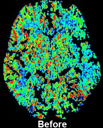
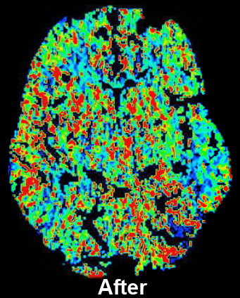
How it works: CT perfusion with acetazolamide (Diamox) consists of a baseline CT perfusion scan of a portion of the brain, followed by injection of acetazolamide (a vasodilatory agent), and then a post acetazolamide CT perfusion scan of the same area. Normally, if blood flow to the brain is decreased, the vessels for that area will expand to maintain adequate oxygen flow, up to a maximum limit. The extent to which this can occur beyond baseline is the cerebrovascular reserve. CT Perfusion with acetazolamide allows assessment of the cerebrovascular reserve.
Equipment: Perfusion studies are usually performed on a Philips 64 or 256 channel CT scanner. OHSU is an ACR accredited CT facility.
Benefits: The study is used to evaluate cerebrovascular reserve, which can help determine the risk of future stroke. The study is often used to help determine which patients might benefit from interventions designed to increase blood flow beyond a very narrow or occluded vessel.
Exam Preparation: Exam preparation is the same as for a CT perfusion study, with additional screening for any potential contraindications to acetazolamide. Additional lab work of sodium and potassium levels are necessary if not performed within two weeks. Potential contraindications to acetazolamide include: Sulfa/sulonamides, severe renal dysfunction, cirrhosis/severe liver disease, adrenocortical insufficiency, hyponatremia, and hypokalemia. A nurse or technologist will interview you for contraindications, and an IV will be placed. Usually labs will be needed prior to the study.
What to Expect: A baseline perfusion scan is performed, followed by injection of acetazolamide over several minutes. It takes about 15 minutes for acetazolamide to have its maximal effect. A post acetazolimde perfusion scan is performed about 15 minutes after injection. Because of this time between scans and the additional time necessary for screening and possible lab work, the amount of time spent in the department will likely be between 1 and 2 hours.
Severe reactions to a single dose of acetazolamide are rare; however, more common reactions that are usually self limited can include brief numbness around the mouth, paresthesias, malaise, and headache.
Adverse reactions to contrast materials are uncommon, but can range from mild to severe. Severe reactions very uncommon. Further information about the risks and benefits of x-rays and contrast material can be found here.
Content by Dr. Louis P. Riccelli
References:

How it works: Arterial Spin Labeling or ASL is a non-invasive method of measuring cerebral blood flow, or blood flow in the brain. The technique does not require intravenous contrast and can be performed on patients of all ages. The scanner creates an image showing relative changes in cerebral perfusion. Additional techniques can be performed that can show actual flow over time (Multiphase ASL). These techniques are most beneficial to patients with vascular stenosis, stroke, arteriovenous malformations, and tumors.
Equipment: Our MRI suite uses the latest Phillips 3.0 tesla magnet coupled with a state of the art Phillips Clinical ASL software package. Processing is performed automatically as soon as the scan is completed and images are available for immediate interpretation.
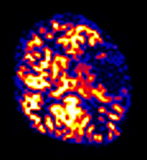
Benefits: ASL is non-invasive so it can be performed on any routine brain MRI on any patient regardless of intravenous access, contrast allergy, or renal status. In young patients ASL is the optimal technique because the child’s veins can be too small to inject a rapid contrast bolus needed for other forms of MRI perfusion (DSC LINK). ASL can show perfusion abnormalities when all other MRI sequences appear normal. The best example of this is a patient with a transient ischemic attack or TIA. These patient’s have brief stroke like symptoms, but may have a normal MRI. ASL perfusion can reveal subtle perfusion changes that could allow possible therapeutic interventions to prevent a future stroke.
Exam Preparation: The technologist will interview you prior to scan to make sure you have no contraindications to being in the MRI scanner. Patients with braces or other metal near the head or neck may not be suitable for ASL because of the artifacts the metal can cause. Also individuals who have received MRI gadolinium based intravenous contrast within 48 hours of the ASL exam may not be suitable for perfusion imaging because of additional artifacts.
What to expect: ASL alone takes approximately 4-5 minutes of scanner time. The study is done in conjunction with routine anatomic imaging.
Content by Dr. Jeffrey Pollock
References
- Pollock JM, Deibler AR, West TG, Burdette JH, Kraft RA, Maldjian JA. Arterial Spin-Labeled Magnetic Resonance Imaging in Hyperperfused Seizure Focus: a Case Report. J Comput Assist Tomogr. 2008 March/April;32(2):291-292
- Deibler AR, Pollock JM, Kraft RA, Tan H, Burdette JH, Maldjian JA. Arterial spin-labeling in routine clinical practice, part 1: technique and artifacts. AJNR Am J Neuroradiol 2008 Aug;29(7):1228-34.
- Deibler AR, Pollock JM, Kraft RA, Tan H, Burdette JH, Maldjian JA. Arterial spin-labeling in routine clinical practice, part 2: hypoperfusion patterns. AJNR Am J Neuroradiol 2008 Aug;29(7):1235-1241
- Pollock JM, Whitlow CT, Deibler AR, Burdette JH, Kraft RA, Tan H, Maldjian JA. Anoxic injury-associated cerebral hyperperfusion identified with arterial spin-labeled MR imaging. AJNR Am J Neuroradiol 2008 Aug;29:1302-1307
- Deibler AR, Pollock JM, Kraft RA, Tan H, Burdette JH, Maldjian JA. Arterial Spin Labeling in Routine Clinical Practice Part 3: Hyperperfusion Patterns. AJNR Am J Neuroradiol. 2008 Sep;29(8):1428-35
- Pollock JM, Deibler AR, Burdette JH, Kraft RA, Tan H, Evans AB, Maldjian JA. Arterial Spin Labeled MRI in Hyperperfused Migraine Evaluation. AJNR Am J Neuroradiol. 2008 Sep;29(8):1494-7
- Pollock JM, Deibler AR, Whitlow CT, Kraft RA, Tan H, Burdette JH, Maldjian JA. Hypercapnia-Induced Cerebral Hyperperfusion: An Underrecognized Clinical Entity. AJNR Am J Neuroradiol. 2009 Feb;30:378-85
- Pollock JM, Whitlow CT, Kraft RA, Tan H, Burdette JH, Maldjian JA. Pulsed Arterial Spin Labeled MRI Evaluation of Tuberous Sclerosis. Am J Neuroradiol. 2009 Apr;30(4):815-2
- Pollock JM, Whitlow CT, Deibler AR, Burdette JH, Kraft RA, Tan H, Maldjian JA. PASL Clinical Applications. MRI Clinics of North America. 2009 May;17(2):315-38.
- Tan H, Maldjian JA, Pollock JM, Burdette JH, Yang LY, Deibler AR, Kraft RA. A Fast, Effective Filtering Method for Improving Clinical Pulsed Arterial Spin Labeling MRI. J Magn Reson Imaging. 2009 2009 May;29(5):1134-9
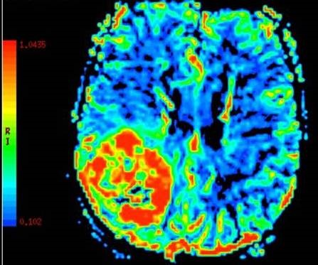
How it works: Dynamic Susceptibility Contrast or DSC is a method of measuring cerebral blood flow, or blood flow to the brain. The technique requires intravenous contrast delivered through an automated power injector attached to an IV. The scanner creates an image showing multiple cerebral perfusion parameters including cerebral blood flow, mean time to enhance, and negative enhancement integral or cerebral blood volume. These techniques are most beneficial to patients with vascular stenosis, stroke, and brain tumors.
Equipment: Our MRI suite uses the latest Phillips 3.0 tesla magnets coupled with a state of the art Phillips Clinical Cerebral perfusion software package. Processing is performed by the technologist as soon as the scan is completed and images are available for immediate interpretation.
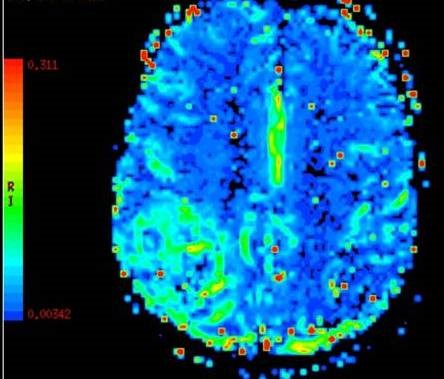
Benefits: DSC perfusion gives the most thorough evaluation of cerebral blood flow. In addition to flow, the technique gives other parameters such as blood volume and transit time that allow the neuroradiologist to more accurately depict the true state of the brain perfusion. These additional parameters have been thoroughly studied with regard to stroke and tumor evaluation.1
Stroke and TIA: The perfusion parameters can show brain tissue at risk of stroke before the stroke has occurred. The best example of this is a patient with a transient ischemic attack or TIA . These patient's have brief stroke like symptoms and but may have a normal MRI. DSC perfusion can reveal subtle perfusion changes that could allow possible therapeutic interventions to prevent a future stroke.
Tumors: Cerebral blood volume correlates with the tumor grade or how malignant the tumor is. Brain tumors are frequently heterogeneous when looked at under the microscope. Perfusion can help guide the neurosurgeon to obtain the most accurate biopsy sample of the tumor to optimize therapy after surgery. Perfusion can also help distinguish a brain metastasis, primary brain tumors, and an infectious process prior to surgery.
Exam Preparation: The technologist will interview you prior to scan to make sure you have no contraindications to being in the MRI scanner. Patients with braces or other metal near the head or neck may not be suitable for DSC because of the artifacts the metal can cause. Patients who have kidney problems may not be able to get MRI contrast without being consented by a radiologist. If you think you have problems with your kidneys please let the technologist and scheduler know. Blood lab values may be obtained prior to the MRI scan to determine if you can receive MRI contrast safely. Patients will be given an IV prior to entering the MRI scanner.
What to expect: DSC perfusion alone takes approximately 2 minutes of scanner time. While the contrast is being injected, the scanner is dynamically acquiring images through the brain. It is very important to remain very still during this portion of the examination. The study is done in conjunction with routine anatomic imaging.
Content by Dr. Jeffrey Pollock
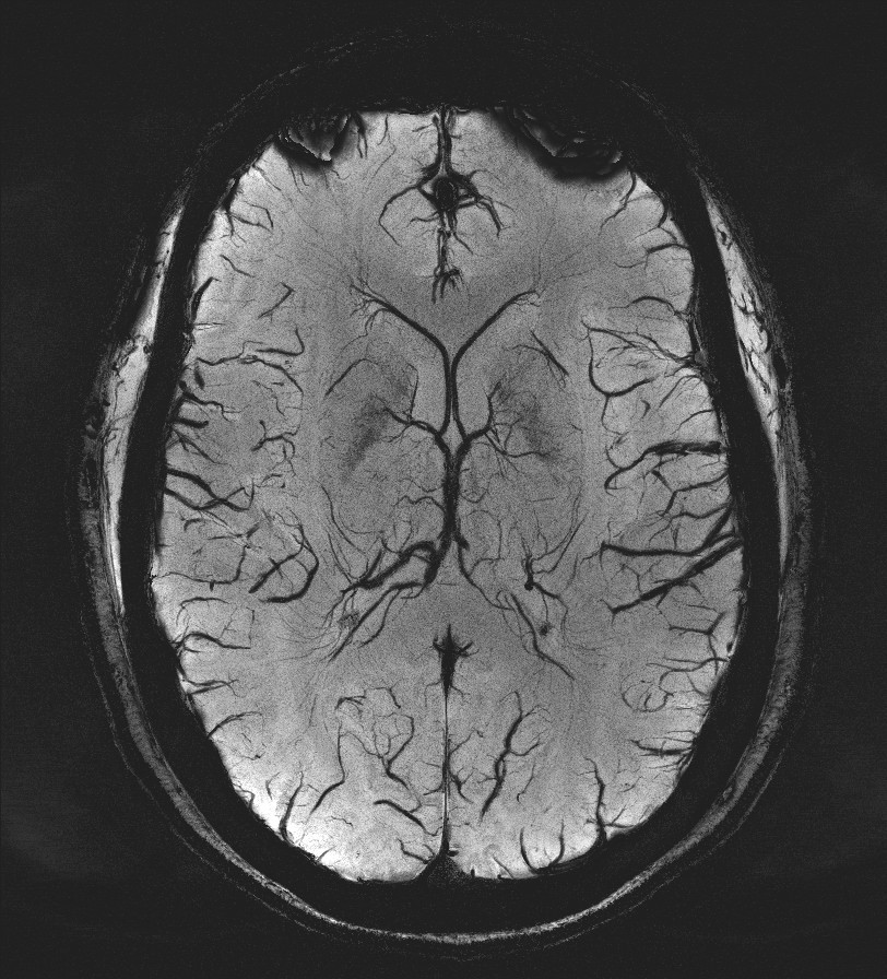
How it works: Susceptibility weighted imaging (SWI) is a non-invasive method of measuring susceptibility in the brain. The technique uses phase and magnitude information to generate an image which reflects the small changes in the local magnetic field. Certain substances such as iron or calcium cause local changes in the magnetic field which can be detected with great sensitivity. The technique does not require intravenous contrast and can be performed on patients of all ages. These techniques are most beneficial to patients with hemorrhages, trauma, concussion, amyloid angiopathy, venous thrombosis, vascular malformations, and tumors.
Equipment: Our MRI suite uses the latest Phillips 3.0 tesla magnets. Processing is performed automatically as soon as the scan is completed and images are available for immediate interpretation.
Benefits: SWI is non-invasive so it can be performed on any routine brain MRI on any patient regardless of intravenous access, contrast allergy, or renal status. Most OHSU MRI exams incorporate some form of susceptibility imaging including Gradient Recalled Echo (GRE), B0, or SWI. SWI is more sensitive than GRE imaging or the B0 images to detect small micro hemorrhages and small vessels. SWI is also able to determine if a structure is composed of calcium or blood based on the phase and magnitude information derived from the SWI.
Exam Preparation: The technologist will interview you prior to scan to make sure you have no contraindications to being in the MRI scanner. Patients with braces or other metal near the head or neck may not be suitable for SWI because of the artifacts the metal can cause.
What to expect: SWI alone takes approximately 5 minutes of scanner time. The study is done in conjunction with routine anatomic imaging. If you are specifically interested in obtaining this image sequence as part of your exam, discuss with your ordering provider as this sequence is routinely included with some protocols, but may be added upon physician request to any MRI protocol at OHSU.
Created by Dr. Jeffrey Pollock
References
- Mittal S, WU Z, Neelavalli J, Haacke EM. Susceptibility-Weighted Imaging: Technical Aspects and Clinical Applications Part 2. American Journal of Neuroradiology. 2009 February; 30(2) 232-252
- Mittal S, WU Z, Neelavalli J, Haacke EM et al. Susceptibility-Weighted Imaging: Technical Aspects and Clinical Applications Part 1. American Journal of Neuroradiology. 2009 January; 30(1) 19-30
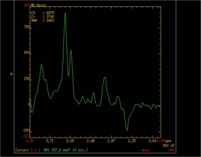
How it works: MR Spectroscopy uses the chemical makeup of a lesion to distinguish it from other possibilities in a differential diagnosis. Each disease process causes different chemical changes in the brain that MRI spectroscopy can detect and identify. MRI spectroscopy can be done in a single point (single voxel) or over a region (multi-voxel). The multi-voxel technique allows the MRI scanner to make an image from the spectroscopy data. The radiologist will select the areas of interest during the examination.
Equipment: Our MRI suite uses the latest Phillips 3.0 tesla magnets coupled with a state of the art Phillips MR Spectroscopy software package. Processing is performed by the technologist as soon as the scan is completed and images are available for immediate interpretation.
Benefits: MRI spectroscopy is most beneficial to patients with suspected tumors or infections. MR spectroscopy can distinguish tumors and infections from different pathologies that can mimic a tumor and it can estimate how malignant a tumor is before surgery. Additionally spectroscopy can identify the regions of the mass that will give the highest yield at surgical biopsy.
Infections: Some intracranial infections can mimic brain tumors. The MR spectroscopy of an infection is very different from a tumor and the two processes can be distinguished.
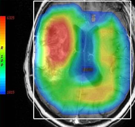
Tumors: The tumor grade can be estimated and possible regions of tumor necrosis can be identified. By knowing the possible grade of the tumor before surgery, the surgeon has the information needed to make the appropriate resection or biopsy. Spectroscopy can also guide the biopsy to the region of the tumor with the most pathology to give the most accurate tumor grade which can affect therapy after resection.
Exam Preparation: The technologist will interview you prior to scan to make sure you have no contraindications to being in the MRI scanner. Patients with braces or other metal near the head or neck may not be suitable for MRI spectroscopy because of the artifacts the metal can cause.
What to expect: DSC perfusion alone takes approximately 5 minutes of scanner time. It is very important to remain very still during this portion of the examination. The study is done in conjunction with routine anatomic imaging.
Created by Dr. Jeffrey Pollock
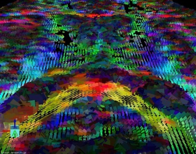
How it works:The white matter is the wiring of your brain. This wiring delivers the electrical signals from your cortex to your extremities. Diffusion Tensor imaging or DTI, uses the magnetic properties of water to create a map of the white matter in the brain. This data can be modeled into a 3 dimensional map of the white matter called tractography.
Equipment: Our MRI suite uses the latest Phillips 3.0 tesla magnet coupled with a state of the art Dynasuite post processing system. Our exams are personally tailored and monitored by a CAQ board certified neuroradiologist.
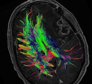
Benefits: DTI is used for presurgical and preradiation planning for an intracranial lesion that could potentially involve eloquent cerebral cortex. The DTI exam is tailored to the location of the lesion and the normal brain adjacent to it. The goal is to allow the neurosurgeon to plan the optimal, least invasive approach to a lesion, and to allow the surgeon to more completely excise the mass or lesion while preserving as much normal function as possible. DTI also allows a patient to better understand the potential risks of having surgery. Our neurosurgical colleagues at OHSU integrate the DTI information into the operating suite using a 3D stereotactic software so they can see the location of the white matter tracts during the surgery. Many studies have shown improved morbidity and increased survival rates in patients who have had preoperative DTI.
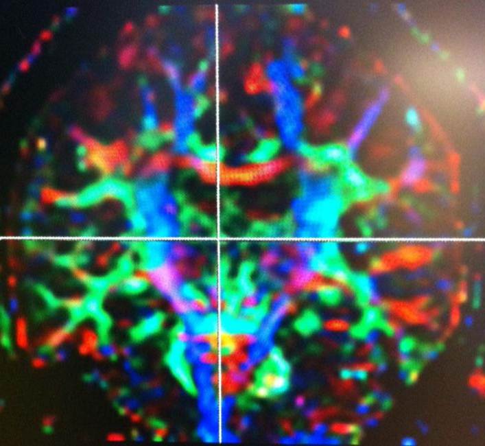
Exam Preparation: The technologist will interview you prior to scan to make sure you have no contraindications to being in the MRI scanner. Patients with braces or other metal near the head or neck may not be suitable for DTI because of the artifacts the metal can cause. The exam is usually done in conjunction with a functional MRI (FMRI).
What to expect: DTI alone takes approximately 2-6 minutes of scanner time. During this time it is very important to remain still. The post-processing of the data by the neuroradiologist can take up to 1 hour. The DTI is usually done along with an FMRI examination.
Content by Dr. Jeffrey Pollock
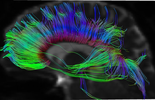
How it works: The white matter is the wiring of your brain. This wiring delivers the electrical signals from your cortex to your extremities. Diffusion Tensor imaging or DTI, uses the magnetic properties of water to create a map of the white matter in the brain. This data can be modeled into a 3 dimensional map of the white matter called tractography.
Equipment: Our MRI suite uses the latest Phillips 3.0 tesla magnet coupled with a state of the art post processing system. Our exams are personally tailored and monitored by a CAQ board certified neuroradiologist.
Benefits: DTI is used for presurgical and preradiation planning for an intracranial lesion that could potentially involve eloquent cerebral cortex. The DTI exam is tailored to the location of the lesion and the normal brain adjacent to it. The goal is to allow the neurosurgeon to plan the optimal, least invasive approach to a mass, and to allow the surgeon to more completely excise a mass while preserving as much normal function as possible. DTI also allows a patient to better understand the potential risks of having surgery. Our neurosurgical colleagues at OHSU can integrate the Tractography information into the operating suite using a 3D stereotactic software so the surgeons can see the location of the white matter tracts during the surgery. Many studies have shown improved morbidity and increased survival rates in patients who have had preoperative DTI.
Exam Preparation: The technologist will interview you prior to scan to make sure you have no contraindications to being in the MRI scanner. Patients with braces or other metal near the head or neck may not be suitable for DTI because of the artifacts the metal can cause. The exam is usually done in conjunction with a functional MRI (FMRI).
What to expect: DTI alone takes approximately 6 minutes of scanner time. During this time it is very important to remain still. The post-processing of the data to generate the trachtography by the neuroradiologist can take up to 1 hour. The DTI is usually done along with an FMRI examination.
Created by Dr. Jeffrey Pollock

How it works: Functional MRI is based on changes in blood flow to regions of the brain associated with a activity. For example, when you move your fingers, the neurons in your brain that tell your finger to move need more oxygenated blood and the body delivers more blood to these cells. The MRI scanner can measure these very small changes in blood flow and display them on an image.
Equipment: Our functional MRI suite uses the latest Phillips 3.0 tesla magnet coupled with a state of the art Invivo paradigm delivery system. Our system uses a 32 inch LCD monitor viewable through a system of mirrors to show patients a customizable visual based FMRI examination. We have technologists who are trained on Functional MRI techniques and all of our exams are personally tailored and monitored by a CAQ board certified neuroradiologist.
Benefits: FMRI is used for presurgical planning for an intracranial operation that could potentially involve eloquent cerebral cortex. The FMRI exam is tailored to the location of the brain lesion, regions of clinical concern, and the normal brain adjacent to it. The goal is to allow the neurosurgeon or clinician to plan the optimal, least invasive approach to a mass, and to allow the surgeon to more completely excise a mass while preserving as much normal function as possible. FMRI allows a patient to better understand the potential risks of having surgery. Many studies have shown improved morbidity and increased survival rates in patients who have had preoperative FMRI.

Exam Preparation: The FMRI is slightly different from other MRI exams:
- You cannot be sedated during the exam. You must be fully alert and responsive. Many patients who are claustrophobic find the LCD monitor a welcome distraction and can still tolerate the examination.
- If you wear corrective lenses for reading, please know your prescription at the time of scheduling. We have MRI compatible glasses and lenses. If you wear contacts, please wear them to the exam.
- Avoid caffeine 1 hour prior to the exam.
- If English is not your primary language please let us know. We currently offer the full FMRI exam in English, Spanish, French, German, Dutch, Italian, and Swedish.
- The language paradigms require the ability to quickly read brief words and sentences of text.
What to expect: FMRI can be done with or without intravenous contrast. If the brain lesion is known to enhance, we prefer to do the study with intravenous contrast to better delineate the lesion. When you arrive the FMRI technologist will review the FMRI tests outside of the scanner. Once in the scanner routine high resolution anatomic images will be obtained first followed by the FMRI. The FMRI exam takes longer than a typical MRI. During this time, (90 minutes) we obtain anatomical images, diffusion tensor imaging (link), perform tractography (link), and the Functional MRI. The functional component of the exam is tailored to the individual patient and may test both language and motor functions. The combination of anatomic imaging, DTI based Tractography, and FMRI gives the clinician the greatest possible amount of information about the functional status of the brain and allows the surgeon to more quickly, accurately, and safely navigate during surgery (combo video link).
Created by Dr. Jeffrey Pollock
Program Faculty
