CT Scan
A CT scan combines X-ray and computer technology to create detailed images of your organs and body structures. Doctors use these images to diagnose your condition or plan treatment.
To get the images, you lie inside a CT scanner. CT stands for computed tomography.
When you have a CT at OHSU, you’ll find:
- Quick scheduling
- Fast results
- State-of-the-art equipment
- Complex scans that aren’t widely available
- A team that puts your safety and comfort first
How CT works
An X-ray beam scans an organ or system from different angles. A computer combines the scans into 3D images.
Before your appointment
We usually schedule you for a 30- to 60-minute appointment. We offer weekday and weekend appointments.
Once your scan is scheduled, you’ll get instructions through MyChart on how to prepare. Depending on your scan, you may need to stop eating and drinking for several hours. Please follow instructions or we may have to reschedule your appointment. If you have any questions, please call us at 503-418-0990.
If you’re anxious about being in the scanner, you can ask your doctor to prescribe medication to help you relax. In that case, please bring someone to drive you home.
Let us know if you:
- Are unable to lie still for more than a few minutes.
- Are pregnant or think you may be pregnant.
- Are breastfeeding.
- Have a kidney condition.
At your appointment
You may be given a substance called contrast by IV or to drink. Contrast helps organs and tissues stand out on imaging. Some people may have a mild allergic reaction to contrast. If this happens, we will assess your symptoms and give you medication if needed. You can ask to do your scan without contrast. We will assess your condition and speak with a doctor about the need for contrast.
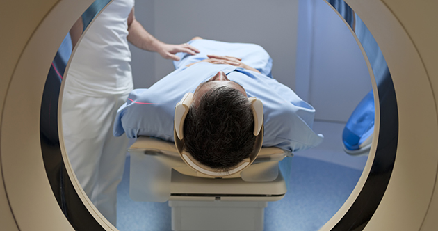
You’ll be scanned while lying on a table that moves in and out of a tunnel-shaped scanner. We have cushions for your comfort.
Scans are painless and take five to 25 minutes. You can talk to us, and we’ll check on you often. If you feel stressed, you can ask to stop. We’ll check the images for quality and see if we need more. We can schedule you for another scan if needed.
After your appointment
You may eat and drink as usual. If you’ve had contrast, drinking extra water will help flush it out.
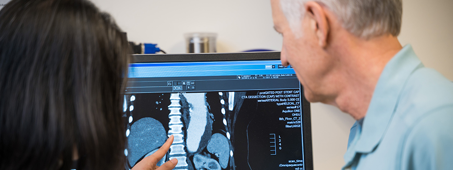
You’ll get your results through MyChart, usually within 24 hours. If you’re having a complex scan, results may take longer because we need more time to process the images.
Types of CT scans
We offer standard and complex CT scans that include:
Cardiac CTA: This checks for coronary artery disease and for any narrowing of the arteries that carry blood to your heart. Learn about the 256-slice CT scan.
Cardiac SPECT CT: Also called dual energy CT, this scan combines CT and nuclear medicine, also called molecular imaging and therapy, to create an image of your heart and how it’s working. Learn more about SPECT CT imaging.
Coronary calcium scan: This checks calcium buildup in arteries that carry blood to your heart. Learn more about coronary calcium scans.
CT perfusion: This checks how well blood is flowing to your brain.
CT TAVR: This checks your heart and vessels. It also checks the aortic valve as part of treatment planning before a heart valve replacement.
Low-dose chest CT for lung cancer screening: This detects lung cancer early in people at higher risk. You may qualify if you are between 50 and 80 years old and are or were a heavy smoker. Learn more about lung cancer screening.
Virtual colonoscopy: Also called CT colonography. This checks for signs of cancer in your large intestine (colon) without sedation or anything being placed inside your body.
Frequently asked questions about CT
How much radiation am I getting?
It depends on the type of scan. Radiation doses range from about 2 to 20 millisieverts (mSv). For comparison, sun exposure and other forms of radiation in the environment expose humans to about 3 mSv each year.
We use the lowest possible dose to get a diagnosis for each patient.
What’s the difference between CT and MRI?
A CT scan uses an X-ray beam that emits a small amount of radiation. A CT is much quicker and quieter than an MRI. A CT is typically better for seeing the structure of bones and organs.
An MRI scan uses magnets and radio waves but not radiation. An MRI takes longer than a CT and can detect things that CT can’t. An MRI is typically better for seeing differences between normal and abnormal tissue.
Which one your doctor recommends will depend on your past imaging, your condition and which scan offers more benefit than risk.
Can a CT scan detect cancer?
Yes. CT scans can show tumors, their size, and whether and where they have spread.
We also use CT scans to assess response to treatment and to look for other anomalies.
Locations
OHSU Diagnostic Imaging Clinic, Marquam Hill (OHSU Hospital)
OHSU Hospital, 10th floor
3181 S.W. Sam Jackson Park Road
Portland, OR 97239
OHSU Diagnostic Imaging Clinic, South Waterfront
Center for Health & Healing, Building 1, third floor
3303 S. Bond Ave.
Portland, OR 97239
OHSU Diagnostic Imaging Clinic, Beaverton
15700 S.W. Greystone Court
Beaverton, OR 97006
Free parking for patients and visitors
Refer a patient
- Refer your patient to OHSU.
- Order diagnostic imaging.
- Call 503-494-4567 to seek provider-to-provider advice.
- Share your imaging.