Message from the Chair
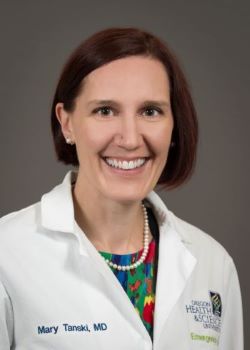
Mary E. Tanski, M.D., M.B.A., Department Chair & Professor
Welcome to the Department of Emergency Medicine at Oregon Health & Science University! The OHSU Emergency Department is a tertiary care referral center, a Level 1 Trauma Center, and an accredited cardiovascular and stroke center. We have a dedicated Pediatric Emergency Department and are a Level 1 Pediatric Trauma Center in collaboration with Doernbecher Children’s Hospital.
Our faculty and staff also provide physician and Advanced Practice Provider staffing for three community hospital partners: Columbia Memorial (Astoria, OR), Hillsboro Medical Center (Hillsboro, OR), and Adventist Medical Center (SE Portland, OR). Together, our team delivers comprehensive, compassionate emergency care for a wide variety of patients across Oregon and beyond.
We are proud to fulfill our three missions: clinical care, education, and research, and I am honored to be part of this passionate, dedicated team.
About the Department
The Department of Emergency Medicine at OHSU is home to 160 clinical faculty members who provide care to approximately 180,000 patients per year across five emergency departments, multiple observation units, and an immediate care virtual program.
- Staff a Level One Trauma ED, Pediatric ED, three community EDs, including one rural access ED, and the Oregon Poison Center
- Ranked 4th in NIH-funded research
- Home to the Center for Policy and Research in Emergency Medicine (CPR-EM)
- Residency Program – Currently trains 33 residents
- Fellowship Programs – Specialized training in Research, Education, Pediatric Emergency Medicine, Administration, Emergency Medical Services (EMS), Toxicology, and Ultrasound
We proudly serve OHSU’s mission of Healing, Teaching, and Discovery.
Education
- Undergraduate Medical Education: Rotations in emergency medicine and subspecialties for OHSU and visiting students
- Residency Program: PGY 1-3 Emergency Medicine Residency Program, established in 1978
- Fellowships: Seven competitive programs in Education, Research, Administration, Toxicology, Ultrasound, Pediatric Emergency Medicine, and Emergency Medical Services (EMS)
- Faculty teach across the School of Medicine, residency program, fellowships, and the Division of Healthcare Management
Oregon Poison Center
- Established by the Oregon Legislature, managed through the DEM
- Board-certified Medical Toxicologists on call 24/7 for complex toxicologic cases
- Hosts learners and provides professional education for healthcare providers statewide
Learn more about the Oregon Poison Center here.
Oregon POLST Registry
- The Oregon POLST (Physician Orders for Life-Sustaining Treatment) Registry helps ensure that patients’ end-of-life care wishes are available to healthcare providers in emergencies.
- DEM faculty and staff participate in education and guidance around POLST forms and their use in emergency care
- POLST forms allow patients to document preferences for resuscitation, medical interventions, and comfort-focused care
- Healthcare providers can access the registry to honor patients’ wishes promptly during emergencies
Research
- Nationally recognized research program; consistently ranked top ten in NIH funding among emergency medicine departments
- Center for Policy and Research in Emergency Medicine (CPR-EM) – Established in 2003, coordinates research and informs health care policy; faculty with backgrounds in medicine, public health, epidemiology, economics, and statistics
Emergency Medicine Simulation
The OHSU Emergency Medicine Simulation program provides hands-on, experiential training for residents, fellows, medical students, and faculty.
- Simulation Labs: State-of-the-art facilities for procedural skills, team training, and critical scenarios
- Interprofessional Education: Collaboration between EM, critical care, nursing, and other healthcare professionals
- Innovative Scenarios: High-fidelity simulations for trauma, resuscitation, toxicology, and disaster response
- Research & Evaluation: Uses simulation to study team performance, clinical outcomes, and best practices in emergency care
Simulation is a cornerstone of OHSU’s training philosophy, preparing learners for real-world emergency and critical care challenges.
Giving / Donations
Support the OHSU Department of Emergency Medicine and help advance patient care, education, and research:
- Contributions help fund residency programs, fellowships, innovative research, and emergency care initiatives
- Donations support faculty development, simulation labs, and patient-centered care programs
- Learn more or give online at the Emergency Medicine Giving page
Your support directly impacts the care we provide to patients across Oregon.
Want to work for the DEM?
Visit the OHSU Jobs page.
Contact Us
If you have an emergent medical condition, do not attempt to contact us on-line. You should go immediately to a hospital or call 911 for emergency transport.
Mailing Address
OHSU Department of Emergency Medicine
Mail Code: CDW-EM
3181 SW Sam Jackson Park Road
Portland, OR 97239
Directions
Turn-by-turn directions, campus maps, and additional information can be found on the Visit OHSU website.