Research at OHSU Dermatology
As part of our department vision, OHSU Dermatology is dedicated to freeing the world of human suffering caused by disorders of the skin. As such, we have made a commitment to aggressively pursue any improvements in patient care, treatment options, and disease prevention through research.
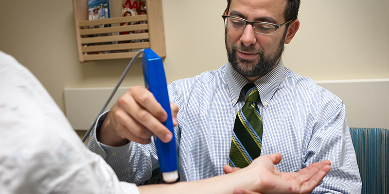
From our basic science laboratories, to translational research, to clinical trials, OHSU Dermatology implements a true bench-to-bedside approach at finding, and implementing advancements to skin health.
Skin condition clinical trials
Explore new treatment options while engaging in research by participating in a clinical trial.
Visit the Skin Conditions Clinical Trials page.
Basic and translational science
Learn about basic and translational science happening across Faculty Labs at OHSU Dermatology.
Visit the Basic and Translational Science page.
Research training program
Learn more about the Molecular Basis of Skin/Mucosa Pathobiology training program at OHSU Dermatology.
Visit the Research Training Program page.