Pathology Services Frequently Asked Questions

Answers about requesting pathology testing services
Explore these frequently asked questions and resources for requesting OHSU Pathology Services. You will find details about:
Frequently asked questions
Submitting a request
-
How do I submit a consultation or service request?
For most services, complete the OHSU Laboratory Testing Requisition Form and submit it with your specimen, slides or blocks to:
OHSU Department of Pathology
Mail Code L471
3181 SW Sam Jackson Park Road
Portland, OR 97239Exceptions include:
- Molecular Genetic Pathology through Knight Diagnostic Laboratories: Visit How to Order at KDL for specific requisition forms and submission instructions.
- Autopsy Services: Contact Pathology Administration directly at 503-494-8276.
- Urgent consults or intraoperative frozen sections: Call 503-494-6776 before sending materials.
-
What information should I include with my request?
For all consultation requests, please include:
- Completed OHSU Laboratory Testing Requisition Form
- Complete clinical history
- Results of relevant imaging studies
- Previous pathology reports, if available
- Referring provider name, institution, phone and fax
- Billing information
- Specimen type and number of blocks, slides or containers submitted
Renal Pathology, Molecular Genetic Pathology and Hematopathology have additional requirements — contact those services before sending.
Specimen and sample preparation
-
How do I prepare and ship tissue specimens or slides?
General preparation guidance by material type:
- FFPE blocks: Ship at room temperature in a padded, rigid container. Include unstained slides, if possible, for IHC or molecular testing.
- Unstained slides: Ship in a slide mailer; include the paraffin block when available.
- Fresh tissue (for renal and molecular studies): Contact the relevant service before sending — requires specific handling and advance coordination.
- Hematopathology fixatives: Contact the Immunology Lab at 503-494-2302 for fixative requirements and technical information.
- Cytology specimens: Contact Surgical Pathology at 503-494-6776 for FNA and fluid submission protocols.
-
What are the renal biopsy submission requirements?
Routine evaluation of a renal biopsy requires specimens prepared for three study types:
- Light microscopy: Formalin-fixed, paraffin-embedded core
- Immunofluorescence microscopy: Fresh or frozen tissue and specific fixative required; contact the lab first
- Electron microscopy: Required for native biopsies; recommended for allografts if the patient has hematuria, proteinuria or other evidence of glomerular disease
Turnaround times and results
-
How long will it take to receive results?
Turnaround times vary by service and case complexity. Estimated ranges:
Anatomic pathology/surgical consultation
- One to two business days
Immunohistochemistry
- One to two business days
Hematopathology (flow cytometry + morphology)
- Two to five business days
Molecular/NGS testing (KDL)
- Comp Heme NGS five to 10 business days
- Solid tumor 10 to 17 business days
- Single gene seven to 10 business days
Check with Knight Diagnostic Laboratories for details about oncology turnaround times.
Renal biopsy (full LM + IF evaluation)
- Next business day (less than 24 hours) for diagnosis
- Clinically urgent rush processing available weekdays and Saturdays
Intraoperative frozen section
- Immediate (during procedure); call in advance
-
How will I receive the pathology report?
Reports are delivered electronically or by fax depending on provider preference. Indicate your preferred delivery method on the request form.
For critical or urgent findings, the pathologist will contact the referring provider directly by phone.
Billing and payment
-
What does pathology consultation cost?
OHSU Pathology provides consultation services at competitive rates. Fees are based on services we perform (surgical pathology interpretation, IHC, molecular testing, etc.). Pricing is available on request. OHSU accepts major insurance carriers. If your patient doesn’t have coverage, they can contact OHSU patient billing and insurance.
-
How do I submit payment or establish a billing account?
Invoices are generated after report delivery. For institutional billing arrangements or to establish a billing account, contact OHSU's pathology billing office. Payment options include check and electronic funds transfer.
Other questions
-
Can community pathologists send cases for second-opinion consultation?
Yes. OHSU pathologists welcome second-opinion consultations from community pathologists, particularly for complex or diagnostically challenging cases. Please submit slides or blocks with a copy of the original report and clinical history. Consultations are accepted throughout Oregon, Southwest Washington and nationally.
-
Do you offer digital pathology or whole slide imaging consultations?
OHSU has whole-slide imaging capabilities through the eSlide Manager and can accommodate digital consultation on select cases. Contact the relevant subspecialty team to discuss digital submission for your case type.
-
How do I request a copy of a previous OHSU pathology report?
Providers may request prior pathology reports through OHSU Health Information Management. Patients may request their own records through MyChart or the OHSU Patient Records office.
Preparing renal pathology specimens1
-
What is an adequate biopsy specimen?
Native kidney biopsy
At least two cores of renal cortex (1 to 2 cm each) are recommended to ensure an adequate sample. When a thin biopsy needle (18 gauge) is used, an additional core is recommended. If an adequate specimen cannot be obtained, a decision will have to be made before the tissue is placed into fixative, as to which one or two of the recommended studies should be requested (see "Dividing the limited specimen" below and/or call one of us to discuss this).
Transplant kidney biopsy
An adequate transplant kidney biopsy to evaluate for rejection consists of at least two needle core segments, each containing renal cortex (see "Dividing the allograft kidney biopsy" below). Avoid:
- Use of forceps (a toothpick or needle is recommended)
- Excessive manipulation
- Drying of the tissue. The tissue should be kept damp in a saline-soaked sponge or filter paper as necessary at the time of the biopsy
- Water (this will cause osmotic damage)
-
How should the unfixed specimen be handled?
Transporting the unfixed specimen
If the specimen is to be transported fresh to your hospital laboratory, it should be placed in a transport medium such as tissue culture medium or moist on a saline-soaked sponge. It should not be placed in water.
The specimen should be divided as soon as possible after the biopsy (preferably, within minutes) before fixation, using a sharp, clean blade. Care should be taken to avoid contaminating the IF specimen with formalin or glutaraldehyde from the blade or other tool. Surgical (wedge) biopsy specimens should be sliced into thin (1-2mm) sections before fixation.
-
How should the unfixed specimen be divided?
Dividing the adequate specimen
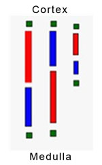
If you use a hand lens or dissecting microscope to distinguish renal cortex from medulla and to identify glomeruli, we recommend the following distribution of tissues:
- Half of the cortex sample in formalin for light microscopy.
- A third of the cortex in Michel's medium for immunofluorescence microscopy.
- A sixth of the cortex in glutaraldehyde for electron microscopy.
If you do not use a hand lens or dissecting microscope, use an empirical method, as detailed by a committee of the Renal Pathology Society, as follows (see reference below):
- Remove a 1 mm segment (green) from each end of every needle core sample. Place these segments into glutaraldehyde for electron microscopy.
- Divide the remaining portions of the cores, alternately 60:40, 40:60, 60:40, etc.
- Place the longer (red) segments in formalin for light microscopy.
- Place the shorter (blue) segments in Michel's or Zeus transport medium for immunofluorescence.
Dividing the limited biopsy specimen
When the biopsy sample is not sufficient to be divided into the three parts as described above, a decision will be necessary as to which one or two of the three recommended studies to request. Consider these guidelines:
- Light microscopy (LM) has highest priority in nearly all circumstances.
- Immunofluorescence microscopy (IF) may have priority in the evaluation of rapidly progressive glomerulonephritis.
- IF is more important than electron microscopy (EM) in most settings of acute renal failure.
- EM is more important than IF in most settings of pediatric nephrotic syndrome.
- IF cannot be performed on tissues fixed for LM or EM.
- The IF tissue can be reprocessed for: Light microscopy (architectural abnormalities can be identified, but cellular artifacts are usually severe) and Electron microscopy (cellular detail is lost but matrix, basement membranes and deposits are usually preserved).
- The LM specimen can be reprocessed for EM with usually satisfactory results.
- Renal medulla is only useful for IF studies in the evaluation of allograft rejection.
Dividing the allograft biopsy specimen
If the allograft biopsy is being done to evaluate for rejection or treatment-related conditions primarily, the cortical sample should be submitted entirely for light microscopy, and a segment of medulla will be sufficient for IF. Electron microscopy is not usually required for evaluation of renal insufficiency in the allograft.
If there is a particular concern about recurrent or de novo glomerular disease in the allograft, the sample should be divided like a native biopsy, with samples of the cortex sent for all three recommended studies (LM, IF and EM).
1 Walker PD, Cavallo T, Bonsib SM and The Ad Hoc Committee on Renal Biopsy Guidelines of the Renal Pathology Society. Practice guidelines for the renal biopsy. Modern Pathology, Vol. 17, Issue 12, Dec. 2004: P1555-1563.
Pathology services directory
Most anatomic pathology consultation requests go through Surgical Pathology. Services with dedicated contacts are listed separately.
- Surgical Pathology (most anatomic pathology services)
- Contact: 503-494-6776
Fax: 503-494-6787
Testing request form - Notes: Bone and Soft Tissue, Breast, Cytopathology, GI/Liver/Pancreas, GU, Gynecologic, Head/Neck/Endocrine, Heart and Lung, IHC, Neuropathology, Pediatric
- Contact: 503-494-6776
- Autopsy Services
- Contact: 503-494-8276
Fax: 503-494-2025 - Notes: Dedicated contact for autopsy requests
Completed reports: Decedent Affairs 503-494-8115
- Contact: 503-494-8276
- Hematopathology (clinical)
- Contact: 503-494-6776
- Notes: For questions and updates on pending cases
- Hematopathology, Immunology Lab
- Contact: 503-494-2302
- Notes: Fixatives and technical information
- IHC Lab (direct)
- Contact: 503-494-5755
- Notes: Direct line for the Immunohistochemistry Laboratory
- Molecular Genetic Pathology, Knight Diagnostic Laboratories
- KDL Client Services
- Notes: Separate ordering system for molecular genetic pathology testing
- Renal Pathology
- Contact: 503-494-6776
- Notes: Contact Renal Pathology for specimen guidelines
Advance notice required before sending fresh tissue
- OHSU Lab Services, Client Services
- Contact: 503-494-7383
1-888-375-4636 (toll free) - Notes: Clinical laboratory testing, test information and lab management
Test directory
Specimen collection information
- Contact: 503-494-7383
Find faculty
Meet our team.
Refer a patient
- Refer your patient to OHSU.
- Call 503-494-4567 to seek provider-to-provider advice.