Magnetic Resonance Imaging Core
About us
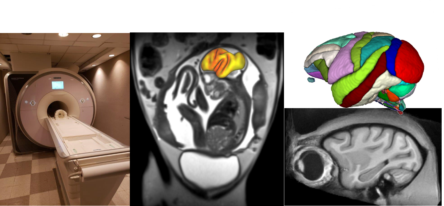
The Magnetic Resonance Imaging (MRI) core provides ONPRC investigators with equipment and assistance to perform MRI exams of NHP subjects. As a satellite facility of the OHSU Advanced Imaging Research Center (AIRC), infrastructure and technical support exist to customize MRI investigations so that researchers can take advantage of the close similarity between human and NHP anatomy and physiology to develop new MRI research and diagnosis techniques and applications.
The ONPRC MRI building is a 2500 sq. ft. facility, housing a Siemens Magnetom 3 T whole-body MR instrument. In the winter of 2017, the MRI system was upgraded with Prisma fit 4G technology. A wide variety of radiofrequency coils are available to support a diverse array of applications. A 16-channel pediatric head coil fits both juvenile and adult rhesus macaque heads. A custom-built 16-channel receive array is also available for studies focused on cerebral cortex. This coil is interfaced with a stereotaxic head positioning frame for the presentation of visual stimuli. Abdominal and fetal imaging is performed using a 15-channel "extremity" coil. Small and large flexible coils are also available that can be shaped to match the contour of the thorax or abdomen of the subjects.
The MRI facility is attached to the Animal Services Building as well as the Primate Multimodal Imaging Center building, to facilitate ease of access for all NHP studies. A comprehensive MRI-compatible anesthesia delivery and physiological monitoring station is used to perform MRI measurements on NHP subjects while monitoring physiological parameters including respiration, arterial oxygenation, pulse rate, non-invasive blood pressure, respiration rate, end-tidal CO2 partial pressure, and ECG.
Contact us
Head, Christopher D. Kroenke, PhD, kroenkec@ohsu.edu
Veterinary technician, Michael Reusz, CVT, reusz@ohsu.edu
MRI scientist, Anahit Grigorian, MS, grigoran@ohsu.edu
MRI Services
-
Brain imaging: Several different options are available for acquiring brain imaging. Whole brain images can be acquired via the 16-channel pediatric head coil. More localized brain imaging can be obtained via the 4 (?) channel special purpose coils, or the (needed description) surface coil.
Abdominal and fetal imaging: a 15-channel knee coil is available for most abdominal scanning requirements, as most NHP subjects will fit into the center of the coil. The spine array in combination with the flex coils available can be used to accommodate larger subjects.
Extremities: Imaging of the extremities is possible through a number of different options, including the flex coils, special purpose coils, and human wrist coil.
Protocols: Generic protocols are already in place for brain and abdominal imaging. The MRI Core staff is available to meet and help design more in-depth imaging protocols to fit study needs.
-
To offer the ability to obtain magnetic resonance data from sedated NHP subjects to the wide pool of ONPRC scientific investigators, the ONPRC MRI core staff are capable of performing a variety of functions.
-
All images acquired on the ONPRC MRI scanner are stored on the OHSU PACs system.
Data retrieval and storage: OHSU Advanced Imaging Resource Center (AIRC) manages all data collected from MRI scanners across OHSU. Here you can find information regarding computing accounts for data processing and storage options.
Physiological Data: The MRI physiological monitor compiles all the data collected during the MRI and formats it into a macro enabled excel spreadsheet. These, along with the data collected by the MRI core staff, are available on the OHSU X drive.
MRI system reservation: Mike Reusz manages a web-based calendar system that ONPRC investigators access to reserve the MRI instrument. An approved institutional animal care and use committee (IACUC) protocol must be referenced in order for investigators to schedule MRI exams. After completion of the experiment, the calendar system is used to charge investigator accounts for use of the MRI instrument.
Safety training: MRI safety training is available for anyone that will be entering the building, and required for anyone that will need badge access to the MRI trailer. There is a compass class available and once completed, a brief tour of the safety features in the ONPRC building is given.
Maintenance of the MRI system: Quality/assurance, management of a calendar system for scheduling the scanner, and billing responsibilities are shared between the ONPRC MRI core, and AIRC staff. These procedures conform to practices developed for the ONPRC MRI system, as well as three other MRI systems located on the OHSU main campus.
-
MRI Rates contact Christopher Kroenke, PhD, kroenkec@ohsu.edu