Integrated Pathology Core
About us
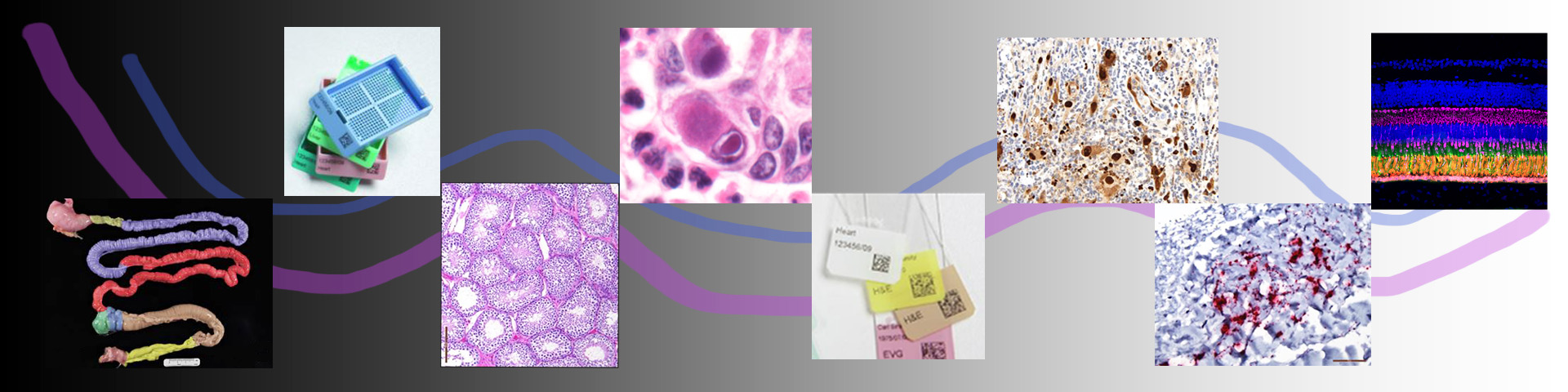
The ONPRC Integrated Pathology Core (IPC), directed by Oleg Varlamov, M.D. Ph.D., provides investigators with state-of-the-art histology and light microscopy imaging and analysis through extensive expertise, services, data analysis, and access to advanced equipment. The core provides routine processing and sectioning of formalin-fixed paraffin-embedded and frozen tissues, special histological stains, next-generation RNAscope/DNAscope in situ hybridization, multiplex immunohistochemistry, digital whole-slide scanning, multi-spectral confocal microscopy, and quantitative image analysis. Imaging and microscopy capabilities include a Leica Stellaris8 spectral confocal (with a white light laser - up to 8 excitation lines from 350 to 790 nm, Acousto-Optical Beam Splitter, Image Navigator module for capturing large tissue areas, resonant scanner, four HyD S detectors, and an Okolab CO2 humidity incubator with a stage top incubator with temperature and gas control for live-cell imaging), a Leica Aperio AT2 high-capacity brightfield whole slide scanner, an Olympus VS120 fluorescence (up to 5 channels; 405, 488, 561, 633, and 750 nm) and brightfield whole slide scanner, and a Keyence BZ-X810 fluorescence microscope. Image analysis is supported by the HALO Image Analysis suite, an advanced quantitative image analysis package that supports unique tissue-specific and disease-specific applications. CellSens (Olympus Bioimaging) and ImageJ are available for basic image analysis. Core personnel are available at every stage of histology and microscopy-based experiments, from planning and training to troubleshooting and quality control.
Contact us
Director, Integrated Pathology Core
Oleg Varlamov, MD, PhD
varlamov@ohsu.edu
503-346-5377
Histology services
Rachel Dannay
dannay@ohsu.edu
503-346-5077
Shipping address
Integrated Pathology Core
Oregon National Primate Research Center
Campus Mail Code L-584
505 NW 185th Avenue
Beaverton, OR 97006
Integrated Pathology Core Services
-
- Pre-project planning
- Expert consultation
- Method documentation
- Publication preparation
-
- Routine and customized tissue processing
- Embedding, sectioning (including large format blocks)
- Routine hematoxylin and eosin staining
- Frozen sectioning
- Custom sectioning: serial, step, large format
- Special histochemical stains
- Cassette and slide printing
-
- Bench top and automated immunohistochemistry
- Antibody optimization
- Multiplex immunohistochemistry
- RNAscope and DNAscope In Situ Hybridization
- Extensive portfolio of antibodies and In situ Hybridization probes validated for use in macaques
-
- Tissue processors
- Leica cassette labeler
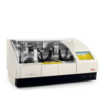
- Leica slide printer, automated cover-slipper, ans auto-stainer
- Semi-automated and manual rotary microtomes
- Biocare IntelliPATH automated immunohistochemistry platform
-
- Leica Aperio AT2 400 slide scanner (bright field)
- Olympus VS120 bright field and fluorescent (up to 5 channels) slide scanner
- Leica Stellaris 8 confocal microscope
-
- HALO Image Analysis: HALO, HALOLink
- Olympus CellSens
- FIJI/ ImageJ