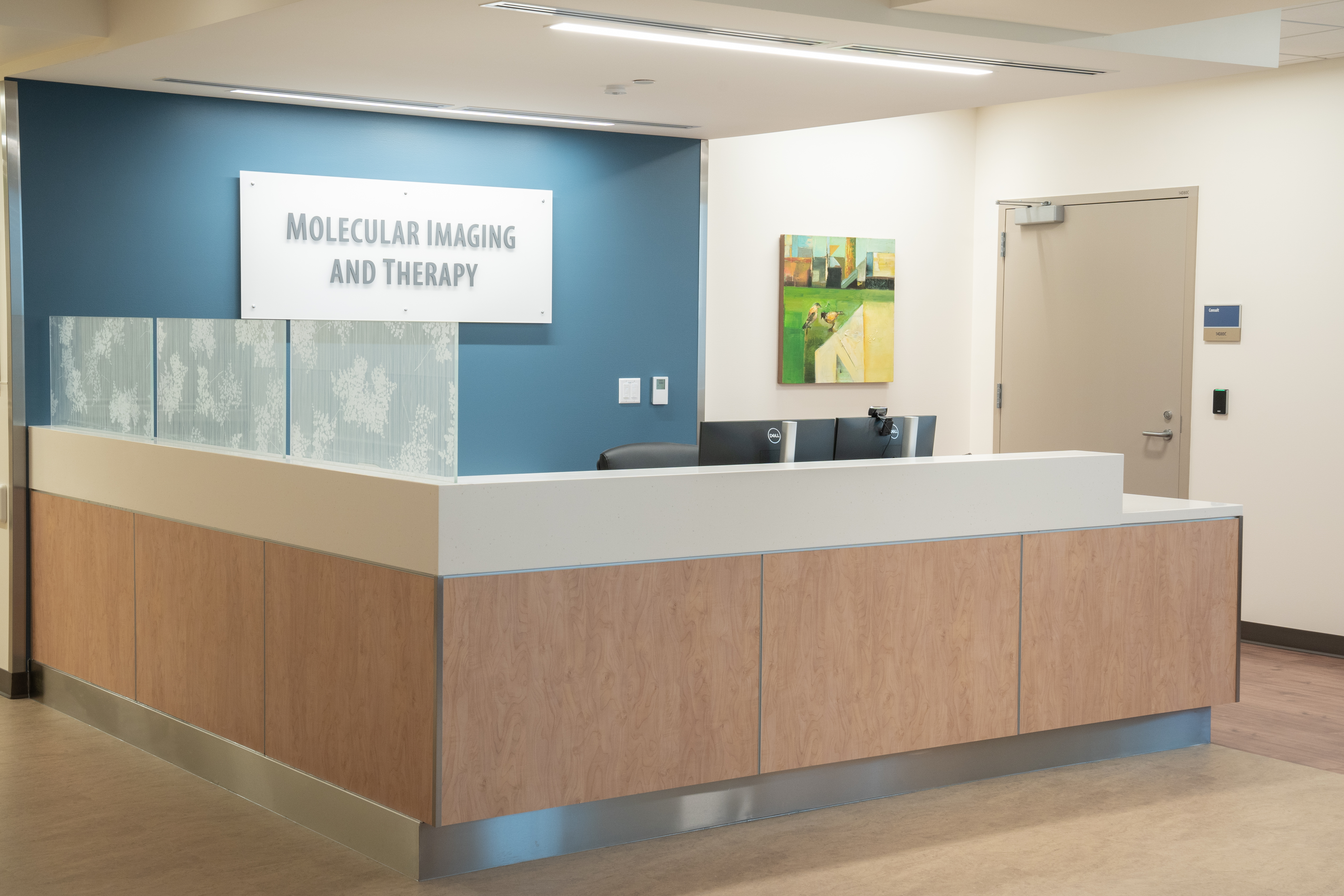
Molecular Imaging and Therapy Clinic
Nuclear Medicine and Molecular Imaging evaluates the function of organs and/or tissues or looks for the presence of disease. It may also be used to follow the progress of treatment of a diagnosis.

At OHSU your care and experience are our number one priority. We are an ACR accredited facility and we take pride in providing the highest quality imaging that you can receive. Our technologists are OBMI certified through the State of Oregon, as well as nationally registered with NMTCB and ARRT. Our board-certified radiologists specialize in Nuclear Medicine. Your appointment is unique to you and each exam is protocoled by one of our Nuclear Medicine Radiologists.
Nuclear Medicine originated with the first Iodine-131 therapy over 77 years ago and is now experiencing a resurgence with several new FDA-approved radiopharmaceuticals for imaging and therapy in the last few years, and the burgeoning field of Theranostics.
Mission Vision Values
Mission
Deliver high-quality molecular imaging and therapy services that provide effective, compassionate care and advocacy for our patients.
Vision
Global leaders in theranostics, utilizing leading-edge technologies to provide advanced and personalized patient care.
Values
• Positivity
• Teamwork
• Integrity
• Compassion
• Wellness
Imaging and Therapy Services Provided
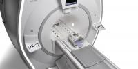
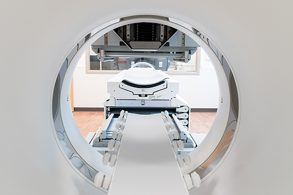
We provide all current FDA-approved Nuclear Medicine imaging services, including gamma (SPECT) and PET for a wide variety of brain, heart, cancer and other indications. We also provide all current FDA-approved radiopharmaceutical therapies including I-131 for thyroid disease, Y90 Sirspheres and Theraspheres for liver cancer, Lu177-DOTATATE (Lutathera) for neuroendocrine tumors, I-131 MIBG (Azedra) for pheochromocytomas and paragangliomas, and Ra223 (Xofigo) and Lu177-PSMA (Pluvicto) for prostate cancer.
Contents
Molecular Imaging and Therapy Clinic
Mission Vision Values
Imaging and Therapy Services Provided
Before the Imaging Procedure
After the Imaging Procedure
Before the Therapy Procedure
After the Therapy Procedure
What is Nuclear Medicine and Molecular Imaging?
Tracer Administration and Image Acquisition
Research
Here at OHSU, we offer the full gamut of diagnostic Nuclear Medicine imaging and therapeutic procedures, participate in many clinical trials, and are developing novel imaging and therapeutic radiopharmaceuticals through the Center for Radiochemistry Research, making us one the leading institutions on the West Coast. For questions regarding your appointment, please call 503-494-8468.
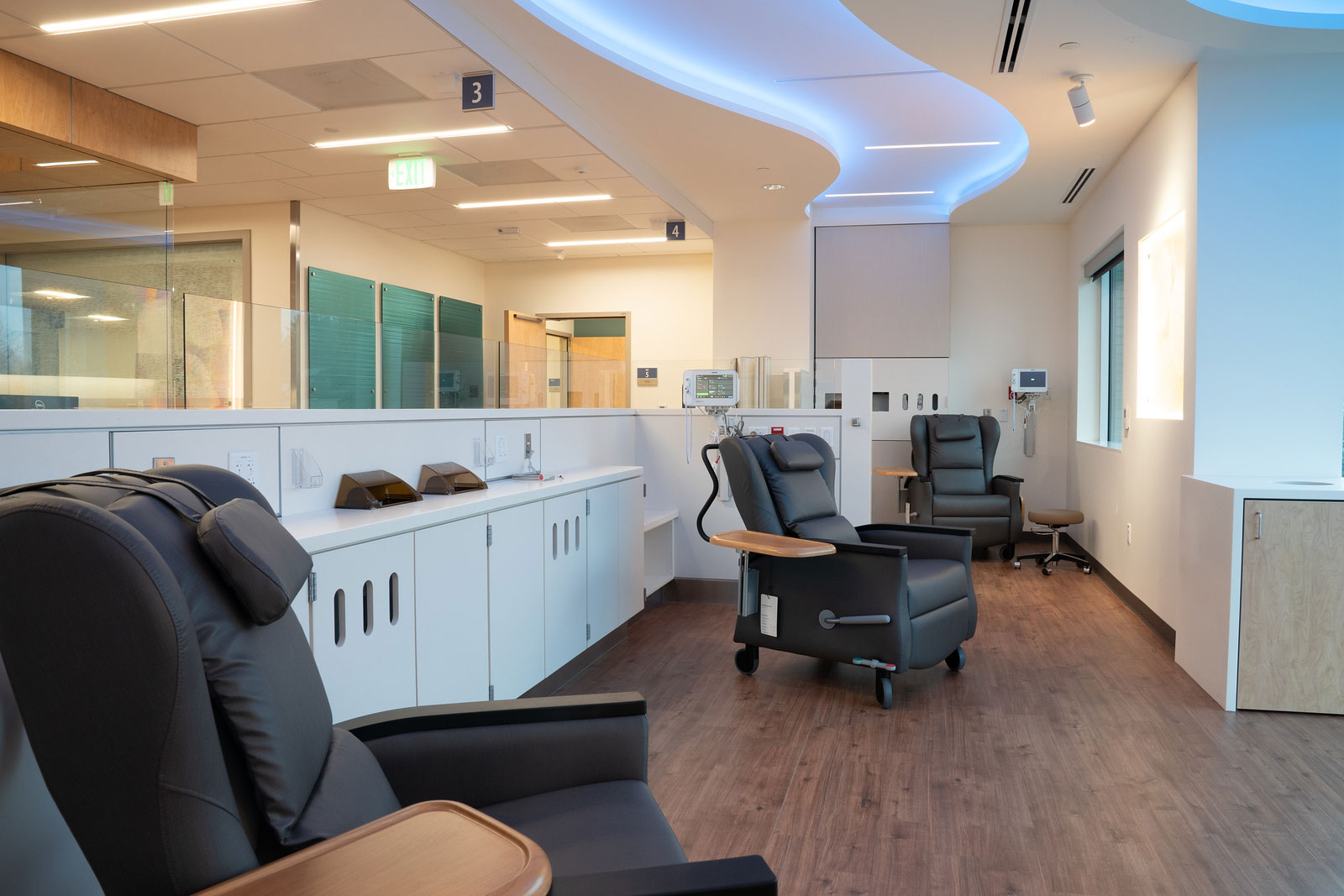
Before the Imaging Procedure
Your appointment is unique to you. Your primary or ordering physician will explain the procedure and offer you the opportunity to ask any questions that you might have about the procedure. A scheduling professional will advise you on any additional steps you will need to take before your exam. You should notify your physician, the radiologist or the technologist if you are allergic and/or sensitive to medications, contrast dyes, or iodine and/or if you are pregnant or suspect you may be pregnant.
What should I expect during my visit?
- A technologist will instruct you on the logistics of your specific exam when you arrive. Our highly trained schedulers can also advise you on how long you will be with us. Generally, nuclear medicine exams follow this process:
- You will be asked to remove any clothing, jewelry, or other objects that may interfere with the scan. If you are asked to remove clothing, you will be given a gown to wear.
- Most likely an intravenous (IV) line will be started in the hand or arm for injection of the radiotracer but the radiotracer may also be administered by inhalation, by oral ingestion, or by direct injection into an organ depending on your diagnosis and the exam ordered.
The scan itself usually causes no pain but for some having to lie still for the length of the procedure might cause some discomfort. The technologist will use all possible comfort measures and complete the procedure as quickly as possible to minimize any discomfort or pain.
After the Imaging Procedure
You will be instructed to drink plenty of fluids and empty your bladder frequently for 24 to 48 hours after the test to help flush the remaining radionuclide from your body. The IV site will be checked for any signs of redness or swelling. If you notice any pain, redness, and/or swelling at the IV site after you return home following your procedure, you should notify your doctor.
Before the Therapy Procedure
Each type of therapy is unique. You will receive a consultation (either in person or virtually based on your preference) from one of our experienced physicians who will guide you through the details of the therapy and discuss your specific case to ensure that you are appropriate for the therapy, have all the information you need to properly prepare for it, know what to expect during it, and how best to plan for afterwards. Throughout the process, you will be assisted by our nurse navigators for all your questions and scheduling requirements.

After the Therapy Procedure
Again, each type of therapy is unique, but in general, after a radiopharmaceutical therapy, you will receive specific radiation safety guidelines to help keep others safe from the effects of the radiation as much as possible. These guidelines will be provided to you in writing and verbally. Also, our nurses and staff are available at any time before or after the therapy to answer your questions about the therapy.
What is Molecular Imaging and Therapy?
Molecular Imaging and Therapy (MIT, also called Nuclear Medicine) is a medical subspecialty that uses small amounts of radioactive isotopes to image and treat various diseases and conditions. The targeted radioactivity is administered internally and the emitted radiation is used to either create images (gamma and PET scans) or treat disease.
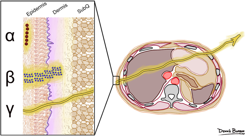
These are the three main types of radiation used in MIT. Each interacts with tissue differently. As such, alpha particles can only be used for therapy, gamma rays are only used for imaging, and beta particles can be used for imaging or therapy.
What is Theranostics?
Theranostics combines the words “therapy” and “diagnostics”, emphasizing the fusion of these approaches within one technology. The goal of theranostics is to personalize and thereby optimize medical treatment by tailoring it to the individual characteristics of a patient, often based on molecular information. While a general term in medicine, the clearest and most common example is in Molecular Imaging and Therapy.
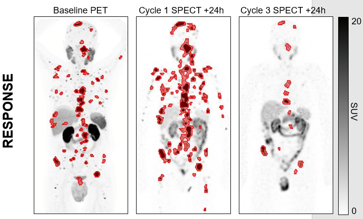
The power of theranostics is shown in the illustration. Everything in red is metastatic disease which is visualized using PET imaging, while the improvement is a result of the therapy, both targeting the same receptor on the cancer cell.
Tracer Administration and Image Acquisition
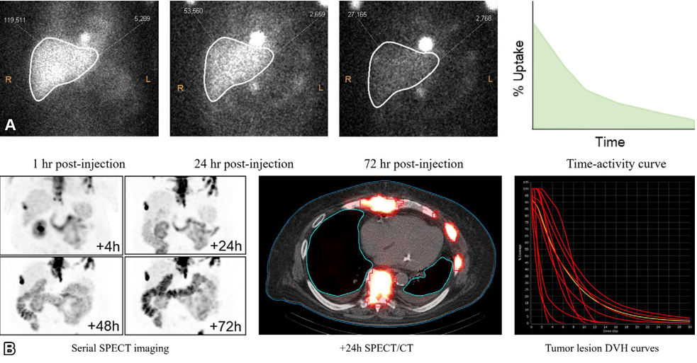
A wide variety of radiotracers are employed in the practice of Nuclear Medicine and Molecular Imaging. They can be injected, inhaled or swallowed, depending on the type of exam being performed. Radiotracers are designed to target specific tissues. For example, in PET scans performed to evaluate tumors, a radioactive form of the glucose molecule (F-18 fluorodeoxyglucose) is injected into the body and is rapidly taken-up by tumor cells that use the glucose for energy. Most radiotracers used have a very short half-life, meaning they exist for only a short period of time before transforming into non-radioactive substances or before being excreted from the body. Most radiotracers used for diagnostic imaging emit a small quantity of radiation that poses minimal risk to your health.
After the radioisotope has been administered and it has collected in the body tissue under study, it emits radiation that is detected by specialized cameras. The amount of time between when a radiotracer is administered and when the images are acquired can range from a few moments to a few days, depending on the specific type of test. The time required to obtain the images may also vary from minutes to several days. Some studies require planar imaging that acquires 2D images.
Based on the theranostics principle, OHSU MIT provides advanced patient specific dosimetry to help optimize the dose and further improve patient outcomes to the therapy.
Research
Our section is engaged in a wide variety of research related to Nuclear Medicine including testing novel radiopharmaceuticals or newer modalities (such as PET/MR), retrospective studies on a number of questions related to existing imaging and therapy we do, and participating in a number of multi-institutional or multi-national prospective studies on the latest developments in molecular imaging or therapy. Some of the current clinical trials we participate in include:
- FaraG: A pilot study using [18F]F‑AraG‑PET imaging to evaluate the immunological response to checkpoint inhibitor therapy (CkIT) in patients with advanced solid tumors
- NeoRay: A Phase I/IIa open-label, multi-center study to evaluate the safety, tolerability, whole-body distribution, radiation dosimetry and anti-tumor activity of [177Lu]-NeoB administered in patients with advanced solid tumors known to overexpress gastrin-releasing peptide receptor (GRPR)
- BED: Prospective study evaluating the role of Axumin® (Fluciclovine or 18F-FACBC) PET in patients with biochemical recurrence of prostate cancer and a negative PSMA PET