MRI Pelvis Hamstring WO MSK Protocol
Scan Notes:
If hardware is present:
- Do Axial and Sagittal STIR instead of fat-sat mid-TE
Last updated: 4/8/19
Charge as: Pelvis WO
Scanner preference: 1.5T or 3T
Coil: Torso Coil
| Plane | Weighting | Mode | Slice | Gap | FAT SAT | FOV | Notes |
|---|---|---|---|---|---|---|---|
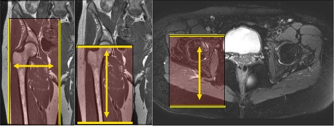
| AXIAL | T1 | TSE | 3mm | 1mm | COVER AREA AS SHOWN BELOW. FOV = Mid hip joint through mid thigh. Slices to cover entire SI Joints to lesser trochanters. | Unilateral | |
| AXIAL | T2 SPAIR | TSE | 3mm | 1mm | SPAIR | COVER AREA AS SHOWN BELOW. FOV = Mid hip joint through mid thigh. Slices to cover entire SI Joints to lesser trochanters. | Unilateral |
| COR | T1 | TSE | 3mm | 1mm | COVER AREA AS SHOWN BELOW. FOV = Sacrum thorough femoral head. Increase slices to cover skin to skin A to P. | Unilateral | |
| COR | STIR (TE=50-60) | TSE | 3mm | 1mm | COVER AREA AS SHOWN BELOW. FOV = Sacrum thorough femoral head. Increase slices to cover skin to skin A to P. | Unilateral | |
| SAG Unilat | T2 SPAIR | TSE | 3mm | 1m | SPAIR | COVER AREA AS SHOWN BELOW. FOV= Iliac crests through lesser trochanters. Slices to cover midline through greater trochanter. | Unilateral |