MR Pfizer A392119 Research Protocol
Scan notes:
- This is a study of the spine and SI joints for Ankylosing Spondylitis. A marker needs to be placed on the patients’ back at the same level as the xiphoid process. The spine is done in two jumps. The two stacks for the total spine need to overlap covering the marker and cover from C1 through S2 (see Fig. 1). If unable to do in two jumps, can do in 3 jumps – change FOV to 300x300 (do not change any other parameters)
- The entire exam is done with the 5CH CTL Spine Coil. Do not use the combo coil or switch to the torso coil for the pelvis.
- For SI Joint Imaging – use the 3 plane loc/midline sag series to get a true mid-sag view of the sacrum. Angle to the posterior aspect of S2 vertebrae, centered A to P on S2 and the third slice of the stack runs along this posterior aspect. (see Fig. 2)
Last updated: 4/12/19
Charge as: Total Spine WO & Pelvis WO
Scanner preference: MR1 only
Coil: Spine coil only
| Sequence | Weighting | Change Parameters? | Change # slices? | Coverage/ Angulation | Notes |
|---|---|---|---|---|---|
| SAG | T1 | NO | NO | Angle to Spine | Upper Station |
| SAG | STIR | NO | NO | Angle to Spine | Upper Station |
| Repeat Reference and Localizer scans. | |||||
| SAG | T1 | NO | NO | Angle to Spine | Lower Station |
| SAG | STIR | NO | NO | Angle to Spine | Lower Station |
| Repeat Reference and Localizer scans. | |||||
| SAG | Survey | NO | NO | Angle to get a True Sag view for Oblique COR planning | |
| SAG | T1 | NO | NO | Angle to posterior aspect of S2 vertebrae (see WWJD and figure 2 for placement instructions) | SI Joints |
| SAG | STIR | NO | NO | Angle to posterior aspect of S2 vertebrae (see WWJD and figure 2 for placement instructions) | SI Joints |
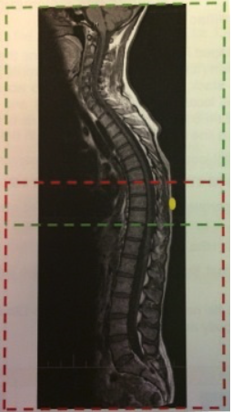
Fig. 1- SAG Stack Planning and Marker Placement

Fig. 2- COR Stack Planning.
NOTE: Blue arrows indicate the posterior surface of S2