MR Female Pelvis WO for FAST Brachytherapy Planning BODY Protocol
Scan Notes
Last updated: 5/11/2022
Charge as Pelvis WO
Scanner preference: MR3
Coil: Torso Coil
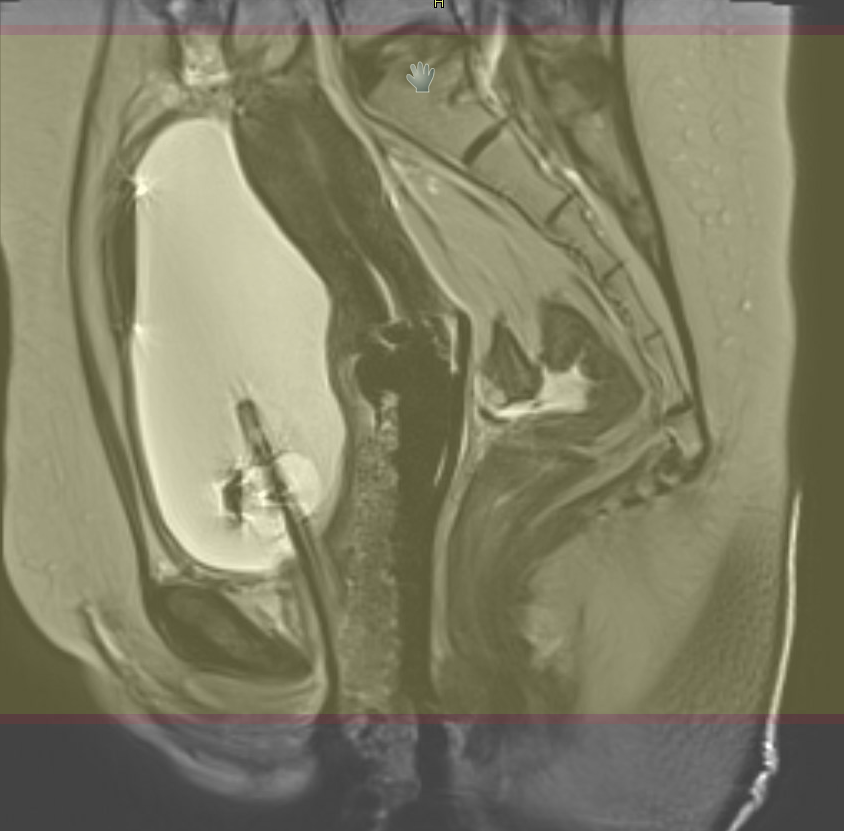
This study is for radiation treatment for endometrial, cervical and vaginal cancer.
- Scan time = Approximately 20 minutes.
- Patient will need to complete a digital or paper screening form before applicator placement to ensure MRI safety.
- Patient will travel to MRI from CT or the OR for this exam.
- Patient will have an applicator in place. The applicator is safe for MRI.
- No vaginal gel is placed for this study.
Post Processing:
- Reformats of 3D T2 at 1 mm to coronal and sagittal planes
| Plane | Mode | S/G (mm) | FS | FOV | Scan Range | Notes | Comments |
|---|---|---|---|---|---|---|---|
| AX T2 | TSE/HI-RES | 3,0 | - | 200-240 mm/ Fit to Patient. Matrix 512 x 256-512. | Entire Vagina, Cervix, or Uterus depending on area of interest | NO ANGLE | NO ANGLE |
| COR T2 | TSE/HI-RES | 3,0 | - | 200-240 mm/ Fit to Patient. Matrix 512 x 256-512. | Entire Vagina, Cervix, or Uterus depending on area of interest | NO ANGLE | NO ANGLE |
| SAG T2 | TSE/HI-RES | 3,0 | - | 200-240 mm/ Fit to Patient. Matrix 512 x 256-512. | Entire Vagina, Cervix, or Uterus depending on area of interest | NO ANGLE | NO ANGLE |
| AX T2 | 3D TSE | 2 mm slice interpolated to 1 mm | - | 270 mm/ Fit to Patient | Cover above the uterus to below the rectum. No need for entire pelvis. | Anterior sat band. Reformat other 2 planes at 1 mm. | Generate 1mm SAG and COR MPRs. Do not reduce slices; SNR is dependent on 3D volume. |