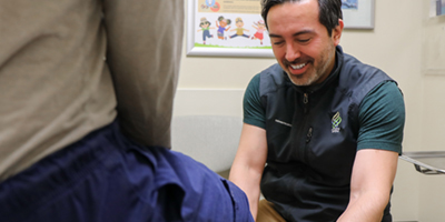
Services we provide
We offer a wide spectrum of diagnostic and therapeutic imaging in an efficient, professional and timely manner.
Welcome message from the Chair
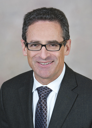
As Chair since 2012, I am proud to be part of the OHSU family, and to lead a department that is increasingly central to the practice and evolution of modern medicine. As a service department, we are dedicated to delivering high quality imaging for our patients, to produce reports for their health care providers that are accurate and informative, and to be readily available for consultation. Our goal is to treat each patient as we would like to be treated ourselves. Providing professional and compassionate care is the expected standard throughout our department.
As OHSU continues to develop as a world class academic medical center, we are committed to advancing the role of imaging in clinical care and research, and to educate and collaborate with the next generation of scientists and physicians. Clinical care, education, and imaging research are the three key components of our work.
As some practical examples, we have introduced and expanded clinical services such as endorectal prostate MRI, in-bore MRI-guided prostate biopsy, MRI-guided high intensity focused ultrasound, advanced molecular imaging including PET/MRI and theranostics, and we are providing critical imaging services to the new Fetal Care and Fetal Surgery Program. We remain part of the ACR registry that tracks and minimizes radiation doses. We are proud of our successful and ongoing efforts to maintain and grow a faculty that is committed to our triple mission and that embodies the institutional values of excellence, diversity, and integrity.
Fergus Coakley, M.D.
Chair of Diagnostic Radiology
Oregon Health & Science University
Radiology CME Grand Rounds and visiting professor series
We invite professors from around the country to speak at our institution to enhance our residents and local radiologists knowledge base. Our visiting lecturers can be viewed online via live streaming in case you are unable to join us in person. Our target audience for our lectures are OHSU, local and regional primary care physicians, specialty physicians, physician associates and nurse practitioners. CME is available. All lectures are held in the Radiology classroom, 10C26, from 12 noon until 1 p.m. unless noted otherwise.