Research on a mission
In the field of ophthalmology, glaucoma has long been a formidable opponent – and a subject of intensive research at Casey Eye Institute. One of the latest basic science projects is led by Casey researcher Benjamin Sivyer, Ph.D., whose laboratory is on a pioneering journey to mitigate glaucoma’s impact on vision.
The challenge of glaucoma
In many cases, elevated intraocular pressure (IOP) in glaucoma leads to an injury to the optic nerve and the subsequent loss of retinal ganglion cells – the sole conduits of visual information from the retina to the brain. Traditional methods of diagnosing glaucoma, such as eye pressure checks and optic nerve examinations, often identify the disease only after damage has occurred. This problem underscores the need for innovative approaches in order to understand glaucoma better and detect it earlier.
Help from our (non-human primate) friends
At the forefront of the Sivyer Lab’s initiatives is the propagation of stem-cell derived retinal ganglion cells in the glaucomatous retinas of non-human primates. “It’s part of an Audacious Goals Initiative (AGI) award we received with several other labs,” Sivyer explains.
The National Eye Institute’s Audacious Goals Initiative for Regenerative Medicine aims to expand vision science and restore vision through cross-disciplinary approaches to retinal regeneration. "Our group is the first in our consortium to implant stem cells to grow human retinal ganglion cells in nonhuman primate tissue," Dr. Sivyer says.
This novel approach involves transforming patient-derived stem cells into retinal ganglion cells and then integrating them into the explanted retinas, aiming to restore the critical connections impaired by glaucoma.
Electrophysiological insights
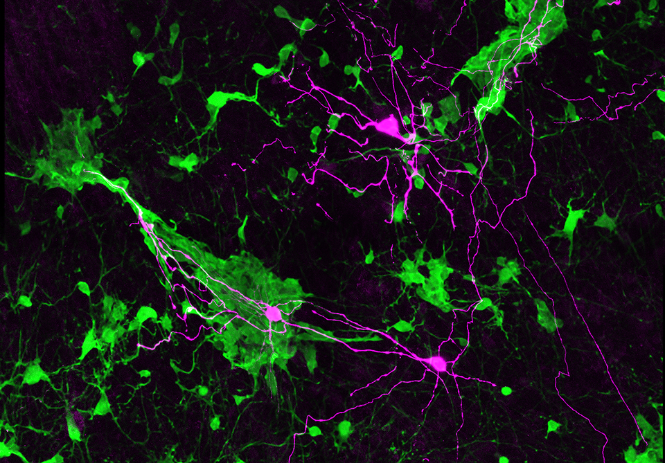
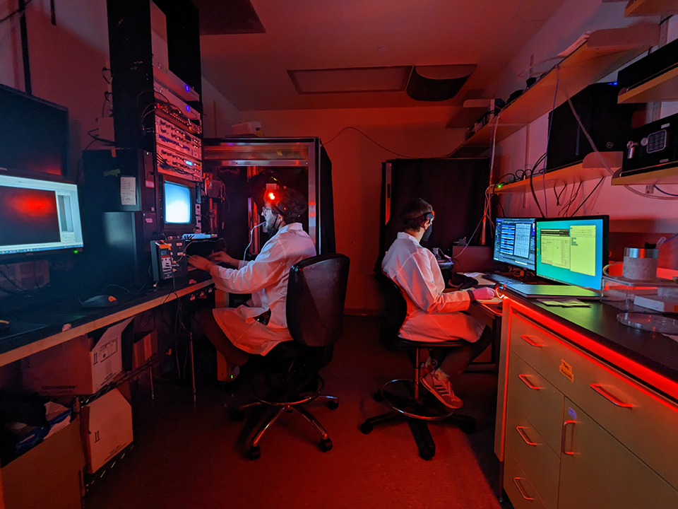
Electrophysiology (EP) reveals how the connections form, explains M.D., Ph.D., student Michael Berry. "We use EP to test how these cells become integrated into the retina. This entails recording from the cells to see if they generate action potentials and are making functional synaptic connections.”
The access to non-human primate tissue – retinas almost identical to humans’ – via the Oregon National Primate Research Center in Beaverton, Ore., and Devers Eye Institute in Portland, Ore., allows high throughput screening of early cell changes in response to glaucoma.
Retinal cells affected by glaucoma do not lose all function right away, Berry says. “Initially, their response to light changes. Studying synaptic function allows us to investigate the changes that cause problems for patients. Eventually, it would be great to identify early on which patients could develop glaucoma, in hopes of intervening earlier and preventing blindness.”
The value of explants
Joe Leffler, Ph.D., a postdoctoral scholar in the Sivyer lab, spends almost all of his time working with the cells themselves. “Using the explant system is exciting because we can test many different variables,” he says. “It’s possible to work on 20 to 30 different conditions at a time, which allows research to proceed at a faster pace." This approach facilitates a deeper understanding of gene expression and cell behavior in glaucomatous conditions.
In addition, Sivyer says, “The recordings Mike and Joe are doing are, I believe, the first example of high density, high quality multi-electrode array recordings from glaucomatous primate retinas.”
Despite its promise, the work is not without obstacles. "So far, the project has been challenging, as we expected. To date, integrating donor ganglion cells in living animals has been problematic," Dr. Sivyer says. “Our group is going back to the drawing board: we are culturing the tissue, using an incubator, adding stem cell-derived ganglion cells, and allowing them to grow and hopefully integrate into the non-human primate retinal tissue.”
Collaboration at OHSU and Casey
The Sivyer laboratory's work is bolstered by collaborations with other research groups at Casey Eye Institute and Oregon Health & Science University (OHSU). “We collaborate with a lot of folks across the university,” Sivyer says. “For example, we have colleagues who are very knowledgeable about synaptic development and lipid nanoparticle delivery,” who can suggest strategies to foster connections between stem cells and retinal tissue.
The science of the future is far from a solo endeavor. As Dr. Sivyer says, “It’s important for science to be teams-based and multi-institutionally based for the greatest benefit, however, working with groups at OHSU and Devers Eye Institute, also in Portland, makes it easy to collaborate."
The environment at Casey Eye Institute fosters collaboration by keeping clinicians and scientists in close proximity – and, of course, by supporting many physician-scientists. Berry says, “Casey is special. Basic scientists asking questions are influenced by physicians who see patients on a day to day basis, and clinically relevant questions and answers arise from that. You’re not siloed from the patients, and this allows us to make the best use of research funding.”
Looking ahead to clinical applications
As the research progresses, insights gained make clinical applications apparent. This work promises to revolutionize both early detection and treatment of glaucoma.
"One of our tasks involves looking at ways to prevent the damage that causes patients to go blind. The other involves a therapy designed for patients at the end of this process, who have lost vision,” explains Berry, underscoring the dual focus on prevention and cure.
These audacious efforts represent a significant leap forward in the fight against glaucoma. The Sivyer lab’s innovative approaches in stem cell propagation, electrophysiological analysis and collaborative research not only enhance our understanding of this complex disease but also pave the way for novel treatments that could significantly improve patient outcomes. As this research progresses, it holds the promise of new hope to those affected by glaucoma – which would mark a new era in eye care.