Nuclear Cardiac Imaging
Cardiac (heart) nuclear imaging is a group of tests that use small amounts of radioactive tracer to create pictures of your heart.
You should know:
- OHSU offers an advanced type of nuclear stress test, which can show how your heart performs when it’s working hard.
- We have a team of heart specialists who can provide diagnoses for complex conditions and offer treatments that aren’t widely available.
- OHSU was the first hospital in the Pacific Northwest to offer PET/MRI, which combines MRI with a PET scan to produce highly detailed images of your heart.
What is nuclear cardiac imaging?
Cardiac nuclear medicine imaging uses tiny amounts of radioactive material to diagnose and sometimes treat heart conditions. The amount of radiation we use is extremely low. It’s not enough radiation to harm your body.
At OHSU, we offer:
- Heart stress test using traditional SPECT imaging
- Special nuclear test (a PYP scan or an HDP SPECT scan) to diagnose cardiac amyloidosis
- Advanced cardiac nuclear stress test, which uses radioactive tracer and an advanced PET scan to create a more detailed picture of your heart and find abnormalities in the blood flow in the heart vessels
- Cardiac SPECT scan, which combines CT technology with nuclear medicine to create a detailed 3D image of your heart
What nuclear imaging is used for
Nuclear cardiac imaging is used to diagnose heart conditions like:
- Coronary artery disease (heart vessel blockages)
- Amyloidosis
- Vascular artery disease
- Hypertrophic cardiomyopathy
- Sarcoidosis
- Inflammation and infection of the heart valves (endocarditis)
What happens during nuclear cardiac imaging?
You will receive detailed instructions on MyChart about how to prepare for nuclear imaging. It’s important to follow these instructions to make sure you are safe and your results are accurate. If you don’t follow the instructions, we may have to reschedule your scan.
At your appointment
When you arrive, our team will ask you to take off items that could disrupt the scan, including jewelry. You’ll get the tracer, usually through an IV in your arm.
Then you’ll lie down on a table that moves in and out of a donut-shaped scanner. The scanner creates images of your heart.
When you’re done with your test, you should drink more water than you usually do for the next 24 to 48 hours. This helps flush the radioactive tracer out of your body.
After your appointment
After your nuclear heart scan, our team will send the results to your doctor. Your doctor will review the results and share their recommendations with you.
Your results play a part in your treatment. But they do not determine your treatment. Your doctor will think about things like your age, other tests, risk factors and overall health in recommending next steps.
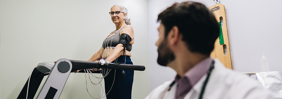
Advanced nuclear stress test
We can use nuclear imaging to do an advanced kind of stress test. This testing helps us see how well your heart functions when it’s working hard.
This test is like a regular exercise stress test except you’ll be injected with a small amount of radioactive tracer. To start, your technician will use an advanced positron emission tomography (PET) scanner to get images of your heart at rest.
Then you’ll get your heart rate up by exercising on a treadmill or an exercise bike. If you can’t exercise, you might get a medicine that can increase your heart work and blood flow. This mimics what happens during exercise.
After your heart rate has increased to the right level, you’ll be scanned again. Your doctor will compare the images to see how well your heart works when it’s under stress.
This advanced nuclear stress test takes about 60 minutes.
SPECT scan
Your doctor might suggest you get a Single Photon Emission Computed Tomography (SPECT) scan. This test combines nuclear medicine with advanced CT images to create a detailed 3D image of your heart.
SPECT can help identify parts of your heart or blood vessels that have reduced blood flow. A SPECT scan can show how well your heart is working. It can also help your doctor find the cause of any pain you have and see if you are having a heart attack.
How does a SPECT scan work?
Before the scan, you'll get radioactive tracer through an IV in your arm. The tracer helps your doctor see how your blood flows.
A computer uses the information from the tracer and combines it with images from the CT part of the scan. It creates a clear and detailed 3D image of your heart that can help your doctor find disease earlier.
This nuclear stress test takes 1 to 2 hours.
Locations
Center for Health & Healing, Building 1, South Waterfront
3303 S. Bond Avenue
Portland, Oregon 97239
Center for Health & Healing, Building 2, South Waterfront
3485 S. Bond Avenue
Portland, Oregon 97239
OHSU Knight Cardiovascular Institute Cardiology Clinic, Beaverton
15700 SW Greystone Ct
Beaverton, 97006
Free parking for patients and visitors
Refer a patient
- Refer your patient to OHSU.
- Call 503-494-4567 to seek provider-to-provider advice.