Center for Radiochemistry Research
The Center for Radiochemistry Research provides scientists with access to a suite of powerful tools for imaging with short-lived radionuclides, data analysis and is integrated with campus centers (SARIC and nuclear medicine clinic) for quantitative imaging of these tracers in vivo that provides spatial and temporal data in order to improve our basic understanding of biochemical pathways, disease processes and help select personalized treatments that enhance clinical care. Highlights of our center include:
- On-campus capacity to develop and produce radiopharmaceuticals to address specific research questions using in-vivo, real-time imaging.
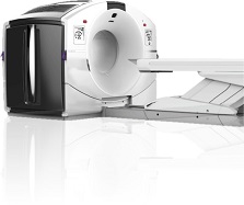
- PET/CT, SPECT/CT (and soon PET/MRI) imaging capability to quantify organ perfusion, assess metabolic pathways, receptor function and density, and measure pharmacodynamics.
- Applications are directed toward a range of models, ranging from small animals to non-human primates, to humans.
What is PET? Positron emission tomography
Positron emission is a radioactive decay process that emits antimatter electrons, called positrons or positive electrons. These positrons are emitted with high velocity but they quickly lose that energy and combine with conventional negative electrons. Then the e+e- pair self-destructs by converting mass to electromagnetic radiation, gamma rays. The gamma rays are always 511 keV and two are emitted in opposite directions. For imaging, we inject a small amount of radioactive biochemical. The radiotracer travels to where the body uses that molecule. From the time of injection to the end of a study, a circle of radiation detectors senses the emitted photon pairs to establish a line along which the radioactive decay event occurred. The decay lines are then reconstructed into a 3D image much as is done for a CT scan. Detection of two gammas simultaneously means that the image detects decay through the body with equal sensitivity, which is essential for quantitative analysis.
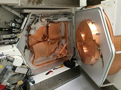
PET tools include a cyclotron to produce short-lived nuclides, dedicated facilities including cleanrooms that contain automated radiosynthesis equipment in hot cells (lead shielded boxes to protect personnel) and a PET scanner to detect radioactivity from positron emission.
For more information
E-mail Hope Phillips at phillhop@ohsu.edu to:
- Schedule a consultation
- Inquire about postdoctoral positions
- Partner on our research