Glaucoma is a sneaky disease. Its damage is subtle and gradual, and may even begin before you notice problems with your eyesight. However, its effects are irreversible and can lead to significant visual disability.
Nearly 3 million American are living with glaucoma, considered a leading cause of permanent blindness worldwide. Although the disease can be detected during a dilated eye exam and managed with medical, laser and surgical treatments, in many cases, those measures may be too little, too late.
While some glaucomas are related to a specific medical condition, how and why the disease develops remains unclear for the majority of cases.
At OHSU Casey Eye Institute, scientists are working together to better understand glaucoma’s underpinnings, gaining insights that will lead to new and more effective ways to diagnose and treat glaucoma before damage occurs.
Their accomplishments are an outgrowth of Casey’s decades of leadership in the field of glaucoma research, which continues to earn international recognition and attract significant funding from the National Institutes of Health, Research to Prevent Blindness and other leading organizations. In October, Casey scientists Ted Acott, Ph.D., John Morrison, M.D., and other glaucoma researchers played prominent roles in a symposium sponsored by the International Society for Eye Research and the Bright Focus Foundation.
“Many patients are affected by the limitations, costs and side effects of our current therapies. This is what drives the collaborations between practitioners and researchers, creating a sense of urgency to address all aspects of the disease,” said Beth Edmunds, M.D., Ph.D., director of Casey’s glaucoma division and associate professor of ophthalmology, OHSU School of Medicine.
Different angles of investigation
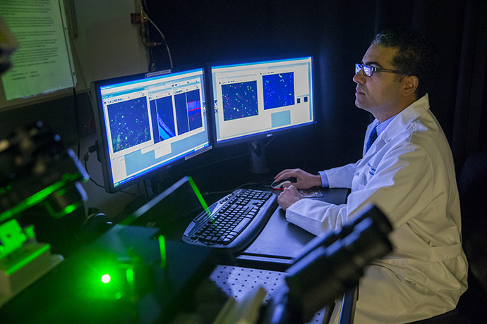
Glaucoma affects the area in front of the lens (anterior) as well as the back of the eye (posterior) and researchers typically focus their work on one of these regions.
At the Lamfrom Biomedical Research Building on the OHSU campus, NIH-supported investigators such as Janice Vranka, Ph.D., are studying the molecular properties that control the way fluid is transported across the trabecular meshwork (TM), a spongy triangle of tissue at the front of the eye. Open-angle glaucoma is the most common form in Western populations and often develops when the fluid inside the eye – called the aqueous humor – is hindered as it flows out through the TM. This obstruction causes a build-up of pressure that over time can harm the optic nerve and cause vision loss.
Shedding light on the inner workings of the TM is key to the development of improved glaucoma therapies, said Vranka, assistant professor of ophthalmology, OHSU School of Medicine.
For Kate Keller, Ph.D., understanding how proteins in the TM regulate eye pressure may lead to new glaucoma treatments. In healthy eyes, TM cells manufacture proteins that aid the flow of the aqueous humor, said Keller, associate professor of ophthalmology, OHSU School of Medicine.
“But in glaucoma, these proteins are slightly different and may cause the outflow channel in the trabecular meshwork to become clogged. Our aim is to look at the differences in these proteins and better understand how they are organized differently than proteins in healthy eyes,” said Keller, whose research was recently published in the journal of Investigative Ophthalmology and Visual Science.
Keller is also collaborating with Mary Wirtz, Ph.D., professor of ophthalmology and molecular & medical genetics, OHSU School of Medicine, whose lab is zeroing in on genes linked to glaucoma. Several years ago, Wirtz identified a gene variant in large families with glaucoma that may contribute to the TM’s proper functioning, said Wirtz, who is teaming up with Keller to learn more about the gene’s interactions and role in regulating eye pressure. “The exciting thing about these discoveries is that they will contribute to better cures that correct this gene mutation.”
Their colleague, Mary Kelley, Ph.D., is also focusing on the TM, adopting a different yet equally intriguing approach. An associate professor of ophthalmology, OHSU School of Medicine, she is the principal investigator of an NIH study examining the role of stem cell therapy in treating glaucoma. In 2017, Kelley won the prestigious Lewis Rudin Glaucoma Prize from the New York Academy of Medicine for showing for the first time that specialized TM cells can be created from stem cells, reintroduced into eyes with glaucoma and correct the underlying cause of the disease.
Much of this impressive work is built on the research of Acott, who for nearly 40 years has made crucial contributions to our understanding of how the aqueous humor is regulated and how this affects glaucoma. Acott, professor of ophthalmology and biochemistry & Molecular Biology, OHSU School of Medicine, was a keynote speaker at the October international symposium, serves on the editorial boards of several leading medical journals and has been recognized for mentoring numerous glaucoma researchers.
Focusing on the optic nerve
“We have a strong research group studying the front of the eye, but that is only part of the story,” said Morrison, explaining that scientists also want to understand the changes that occur in the back of the eye when eye pressure is elevated, especially before glaucoma is detected.
Morrison, whose glaucoma research spans three decades, is joining forces with other Casey investigators studying retinal ganglion cells and their axons, which form the optic nerve and send images to the brain. The optic nerve, composed of approximately a million retinal ganglion cell axons, exits the eye at the optic nerve head, which is the initial site of injury from glaucoma. Morrison, who is professor of ophthalmology, OHSU School of Medicine, is particularly interested in learning how these cells’ genes, and those of the optic nerve head, respond to the accumulating effects of elevated eye pressure and damage the optic nerve.
One of those researchers, Benjamin Sivyer, Ph.D., recently was awarded a major grant from the BrightFocus Foundation to develop more sensitive methods for studying the onset of glaucoma. Sivyer, assistant professor of ophthalmology, OHSU School of Medicine, said his goal is to pinpoint beginning changes to retinal ganglion cells following injury to the optic nerve. “We hope our research will lead to earlier diagnosis of glaucoma,” he said, and more importantly, uncover mechanisms in the retina that will slow or stop the death of these cells.
Physician-scientist Shandiz Tehrani, M.D., Ph.D., associate professor of ophthalmology, OHSU School of Medicine is among the first to repurpose a small molecule drug to prevent optic nerve axons from degenerating. Called fasudil, the drug is approved for use outside the U.S. for stroke and other neuro-degenerative conditions.
“Our early findings showed that fasudil protected optic nerve axons by 50 percent in a small animal model of glaucoma. This was our first clue that a small molecule administered as a drop, injection or pill may directly protect axons against glaucoma,” said Tehrani, whose work is supported by the NIH and Research to Prevent Blindness. Tehrani and his team hope these investigations will eventually pave the way for new neuroprotective glaucoma therapies.