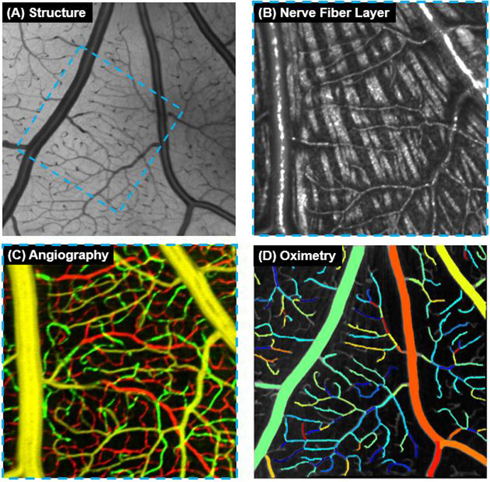
en face images of (A) entire retina and (B) nerve fiber layer slab, (C) angiographical en face image of three
retinal vascular plexuses, and (D) capillary-segment-wise oximetric en face image on SVP. The blue dash boxes
indicate the scan region in (B) and (C). SVP: superficial vascular plexus; ICP: intermediate capillary plexus; DCP:
deep capillary plexus; sO2: oxygen saturation. ©Images courtesy of Dr. Yali Jia.
Innovations: Inside the Lab 2021
Ophthalmic imaging expert Yali Jia, Ph.D. and her team constructed a spectroscopic optical coherence tomography (OCT) instrument using visible-light band. They paired this instrument with a novel algorithm to extract the blood oxygen saturation in retinal capillaries in a preclinical model. Jia’s lab is the first to bring this technology to the capillary level.
“If we can detect vascular oxygenation alteration before patients have vascular morphological changes, it will be a breakthrough as an early marker of disease,” Jia said. “Detection of capillary oxygenation impairment is even more appealing than that of major vessels. This technology has applications for retinal diseases and glaucoma but could also impact imaging for all vascular diseases, including stroke.”
Jia’s lab is now concentrating on realizing this potential tool for clinical use. Compared to conventional methods, OCT oximetry can provide three-dimensional information on specific vessels. It is also efficient, since the data can be processed from the same scans that generate structural OCT and OCT angiography.
Using the same device, Jia’s lab has also worked to generate cellular-level retina images in a preclinical model by improving the hardware sensitivity and software registration accuracy. The images produced by visible-light OCT (vis-OCT) provide finer axial resolution than near-infrared OCT for a given wavelength bandwidth.
“Compared to other imaging modalities with cellular-level resolution, vis-OCT can maintain a depth of focus at a range encompassing nearly the entire retina. Using vis-OCT, these single neurons with different functions at different retina layers can be investigated simultaneously in a 3D volume scan.”
Jia believes vis-OCT could potentially replace traditional histology of tissue staining and collection.
“Vis-OCT is one of the most exciting new frontiers for clinical ophthalmology,” she said. “With superior resolution and the ability to measure capillary oximetry, vis- OCT likely will provide new insights into diseases that lead to blindness and improve our ability to detect and manage these diseases in the earliest stages, before vision is affected.”
Yali Jia, Ph.D. previously developed split-spectrum amplitude-decorrelation angiography (SSADA), a major breakthrough that transitioned OCTA from a purely research technology to the clinic. Her original paper on this subject, published in 2012, has been cited 1350 times. Her recent work investigating novel OCT technologies, including highperformance OCT/OCTA device, the use of artificial intelligence to detect and classify retinal pathologies, spectroscopic OCT to measure capillary oximetry.