Richard G. Weleber Ophthalmic Diagnostic Service

The best techniques to measure eye health
The Richard G. Weleber Ophthalmic Diagnostic Service uses visual electrophysiology and perimetry techniques to measure the health and function of the eye and visual system. As part of the ophthalmic genetics division at OHSU Casey Eye Institute, we offer electrophysiology, full-field ERG, multifocal ERG and perimetry diagnostic testing for patients.
OHSU Casey Eye Institute has a long history of patient testing and care that started with the pioneering work of Dr. Richard Weleber, a clinician-scientist in the field of Ocular Genetics, retinal degenerations, visual electrophysiology and visual field methodologies. In 1974, Dr. Weleber founded the first electrophysiology service at OHSU and was the director of the Visual Function Service for 39 years. Dr. Weleber was involved in the design and conduct of clinical trials, has authored over 200 peer-reviewed publications, and is the recipient of numerous honors and awards.
Testing available
What is a visual electrophysiology test?

The first step in seeing is when a pattern of light from an object is projected on the retina at the back of the eye. The pattern of light is converted by the retina into very small electrical signals and sent to the brain along the optic nerve, where the sensation of “seeing” occurs. Testing these electrical signals is called visual electrophysiology. It can be done non-invasively with minimal risk and it gives information regarding the eye, nerve and brain function that helps the doctor make decisions on diagnosis and treatment.
The test to measure the small electrical signals of the eye is called Electroretinogram (ERG). The photo on the right show the electrode which consists of a thin conductive fiber that contacts the eye and is held in place by the sticky pads. An ERG provides a non-invasive functional test of the retina.
The tests are carried out by Clinical Physiology Technicians. The results for outside testing requests will be interpreted by a consultant electrophysiologist. A report will be sent to the doctor who referred the patient.
What is full-field ERG testing?
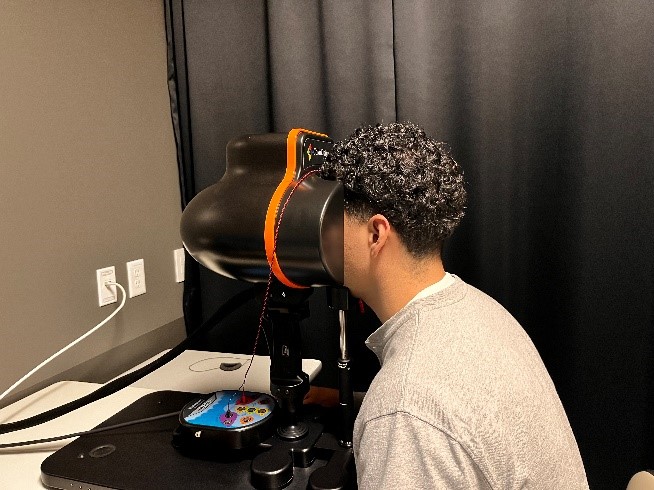
The full-field ERG (ffERG) measures the response of the retina to a luminance stimulus that can be used to separate the function of the cone system from the rod system and the photoreceptors from the inner nuclear layer. Since the ffERG is a massed response it cannot be used to assess localized retinal dysfunction such as central macular cone function.
This chart shows the responses that are recorded from a full-field ERG test. The size, shape and speed of the responses measures the health of the retina.
We use the International Society for Clinical Electrophysiology of Vision (ISCEV) standard testing protocol and the traces above are example waveforms recorded from the corneal surface showing A. Dark adapted (DA) rod response (0.01 cd*s/m2), B. DA mixed rod and cone responses (10 cd*s/m2), C. Light adapted (LA) cone response (3.0 cd*s/m2), D. LA 30Hz flicker cone isolating response. The responses are quantified by measuring waveform amplitude and latencies.
What is multifocal ERG testing?
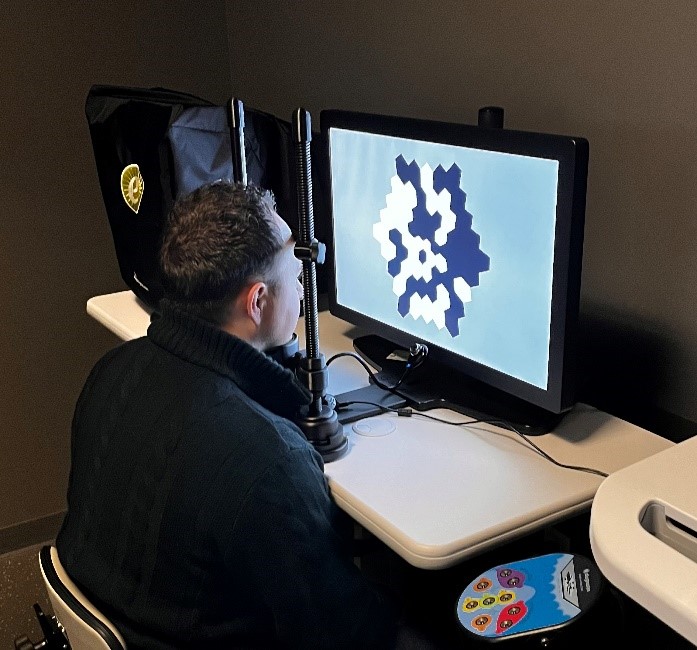
While a ffERG is used to determine for generalized retinal function, a multifocal ERG (mfERG) is useful for mapping discrete areas of retinal function and produces a topographic map of sensitivity. It uses a contrast reversing stimulus that is projected over 30 to 40 degrees of the central visual field and uses a luminance response to test macular cone system function.
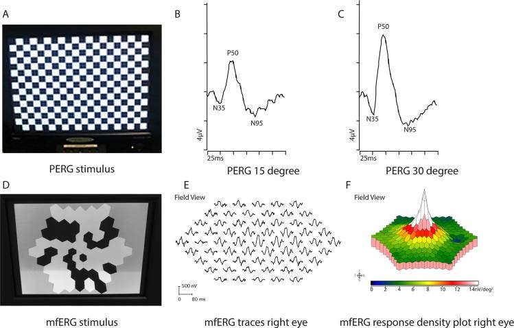
This figure above shows the stimulus projected on computer display for mfERG (left). It consists of a mosaic of hexagons that independently shift between black and white at a rapid rate. Because the subject is required to maintain fixation on a central target, each hexagon stimulates a different location of retina permitting topographic mapping of sensitivity (middle) that is represented by the waveform amplitude. These results can be used to create a 3D map of sensitivity (right). Here, a high central fovea response can be seen. We use the ISCEV standard protocol for mfERG.
What is Perimetry?
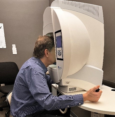
Perimetry is a behavioral test that is used in ophthalmology to assess a patient’s visual function throughout their visual field. It is useful for detecting pathologies and following them over time to determine whether they are progressing or stable. It is also useful for visual ability testing such as testing a person’s ability to drive. The two types of perimetry that are used to assess the visual field sensitivity are static and kinetic perimetry. Kinetic is available to outside referral testing.
Kinetic Perimetry
Kinetic perimetry uses a moving target of a particular size and intensity that passes through the visual field while the subject maintains fixation on a central location in the perimeter. As soon as the stimulus is observed, a button press registers the location within the visual field. In this way, the technique can be used to map the boundaries of central and peripheral visual fields.
The figure shows an example of kinetic visual fields of the left eye (left) and right eye (right). Each colored line is called an isopter and corresponds to a target size. For example, the red and blue lines correspond to the largest target size and can be seen furthest into the periphery. The purple line is the smallest target, hardest to see, and therefore cannot be seen until it encroaches on sensitive area of central retina.
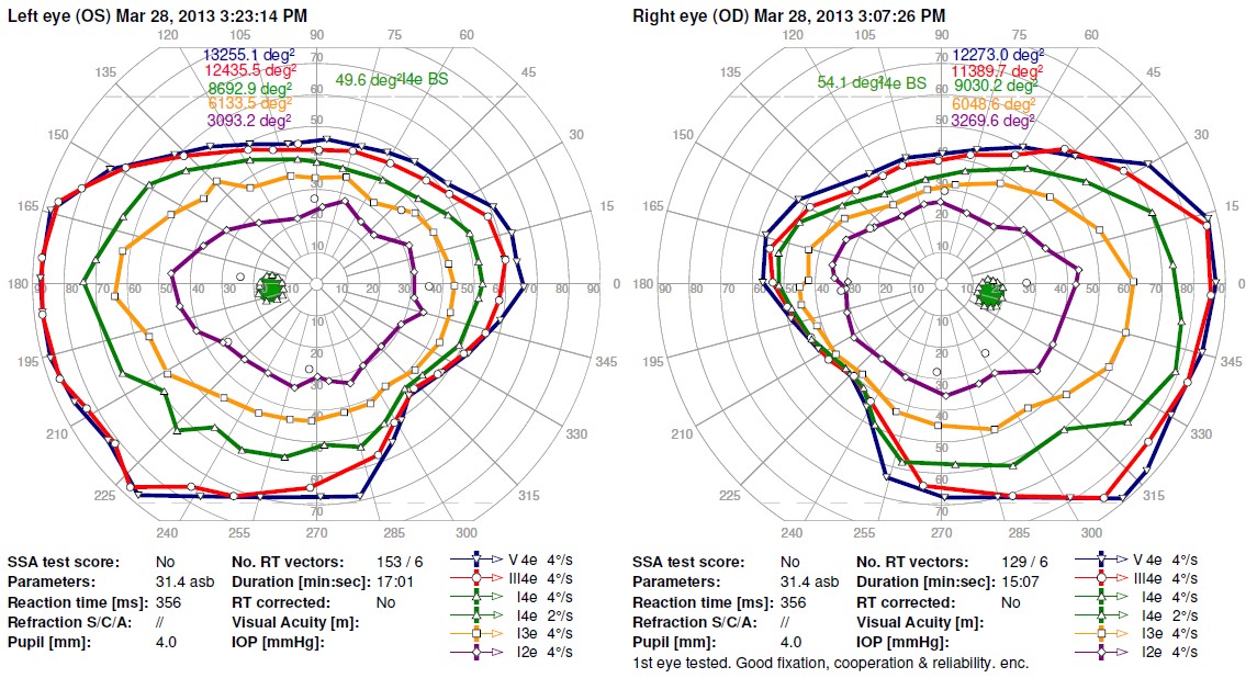
The figure shows an example of kinetic visual fields of the left eye (left) and right eye (right). Each colored line is called an isopter and corresponds to a target size. For example, the red and blue lines correspond to the largest target size and can be seen furthest into the periphery. The purple line is the smallest target, hardest to see, and therefore cannot be seen until it encroaches on sensitive area of central retina.