Renal Pathology Additional Information and FAQ
Consultation information
For fixatives and technical information
- Call the Immunohistochemistry Laboratory at 503-494-5755, or
- FAX us at 503-494-6787 (Attn: Immunohistochemistry Lab)
For general information and assistance
- Call the Surgical Pathology Office at 503-494-6776, or
- FAX us at 503-494-6787 (Attn: Surgical Pathology)
Reports
Preliminary findings are based on the light microscopy findings usually provided by phone to the referring nephrologist or pathologist. A written preliminary report (pending electron microscopy) is available on request. Written final reports are provided for all consultations. Contact us to arrange to have your reports automatically faxed to a secure (office) site after it is signed.
The fixatives
The recommended fixatives are listed below. Please contact us if you would like us to supply these fixatives.
- For light microscopy (LM): 10% aqueous formaldehyde (formalin). This is readily available in most pathology laboratories. Paraformaldehyde and Carnoy's fixative are also satisfactory.
- For Immunofluorescence (IF) Microscopy: Michel's fixative (Modified Michel's Tissue Fixative - Wampole Laboratories, Cranbury, N.J.).
- Electron Microscopy (EM): 2-3% glutaraldehyde. Paraformaldehyde is also suitable
Billing and charges
The following is a list of the usual services and CPT codes for evaluation of a renal biopsy:
| Service | CPT Code | # of Units |
|---|---|---|
| Light microscopy (for tissue processed in our laboratory) | 88305 | 1 |
| Special Stains | 88313 | 3 |
| Immunofluorescence microscopy | 88346 | 1 |
| Immunofluorescence microscopy | 88350 | 7 |
| Electron microscopy | 88348 | 1 |
| Outside consultation (outside slides) | 88321 | 1 |
For technical pricing please call 503-494-6775. For professional pricing please call 503-494-6010.
Frequently asked questions
-
Native kidney biopsy
At least two cores of renal cortex (1 to 2 cm each) are recommended to assure an adequate sample. When a thin biopsy needle (18 gauge) is used, an additional core is recommended. If an adequate specimen cannot be obtained, a decision will have to be made before the tissue is placed into fixative, as to which one or two of the recommended studies should be requested (see "Dividing the limited specimen" below and/or call one of us to discuss this).
Transplant kidney biopsy
An adequate transplant kidney biopsy to evaluate for rejection consists of at least two needle core segments, each containing renal cortex (see "Dividing the allograft kidney biopsy" below).
-
Avoid
- Use of forceps (a toothpick or needle is recommended).
- Excessive manipulation.
- Drying of the tissue. The tissue should be kept damp in a saline-soaked sponge or filter paper as necessary at the time of the biopsy.
- Water (this will cause osmotic damage).
Transporting the unfixed specimen
If the specimen is to be transported fresh to your hospital laboratory, it should be placed in a transport medium such as tissue culture medium or moist on a saline-soaked sponge. It should not be placed in water.
-
The specimen should be divided as soon as possible after the biopsy (preferably, within minutes) before fixation, using a sharp, clean blade. Particular care should be taken to avoid contaminating the IF specimen with formalin or glutaraldehyde from the blade or other tool. Surgical (wedge) biopsy specimens should be sliced into thin (1-2mm) sections before fixation. Guidelines are offered below for dividing the specimen.
Dividing the adequate specimen
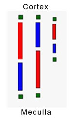
If you use a hand lens or dissecting microscope to distinguish renal cortex from medulla and to identify glomeruli, we recommend the following distribution of tissues:
- 1/2 of the cortex sample in formalin for light microscopy.
- 1/3rd of the cortex in Michel's medium for immunofluorescence microscopy.
- 1/6th of the cortex in glutaraldehyde for electron microscopy.
If you do not use a hand lens or dissecting microscope, an empirical method is recommended, as detailed by a committee of the Renal Pathology Society, as follows (see reference below):
- Remove a 1 mm segment (green) from each end of every needle core sample. Place these segments into glutaraldehyde for electron microscopy.
- Divide the remaining portions of the cores, alternately 60:40, 40:60, 60:40, etc.
- Place the longer (red) segments in formalin for light microscopy.
- Place the shorter (blue) segments in Michel's or Zeus transport medium for immunofluorescence.
Dividing the limited biopsy specimen
When the biopsy sample is not sufficient to be divided into the three parts as described above, a decision will be necessary as to which one or two of the three recommended studies to request. The following comments are offered to help guide your judgment:
- Light microscopy (LM) has highest priority in nearly all circumstances.
- Immunofluorescence microscopy (IF) may have priority in the evaluation of rapidly progressive glomerulonephritis.
- IF is more important than electron microscopy (EM) in most settings of acute renal failure.
- EM is more important than IF in most settings of pediatric nephrotic syndrome.
- IF cannot be performed on tissues fixed for LM or EM.
- The IF tissue can be reprocessed for: Light microscopy (architectural abnormalities can be identified, but cellular artifacts are usually severe) and Electron microscopy (cellular detail is lost but matrix, basement membranes and deposits are usually preserved).
- The LM specimen can be reprocessed for EM with usually satisfactory results.
- Renal medulla is only useful for IF studies in the evaluation of allograft rejection.
Dividing the allograft biopsy specimen
If the allograft biopsy is being done to evaluate for rejection or treatment-related conditions primarily, the cortical sample should be submitted entirely for light microscopy, and a segment of medulla will be sufficient for IF. Electron microscopy is not usually required for evaluation of renal insufficiency in the allograft.
If there is a particular concern about recurrent or de novo glomerular disease in the allograft, the sample should be divided like a native biopsy, with samples of the cortex sent for all three recommended studies (LM, IF and EM).
Reference
Walker PD, Cavallo T and Bonsib SM: Practice guidelines for the renal biopsy. Modern Pathology 17:1555-1563, 2004.