PET/MRI: Advancing Patient Care and Accelerating Discovery
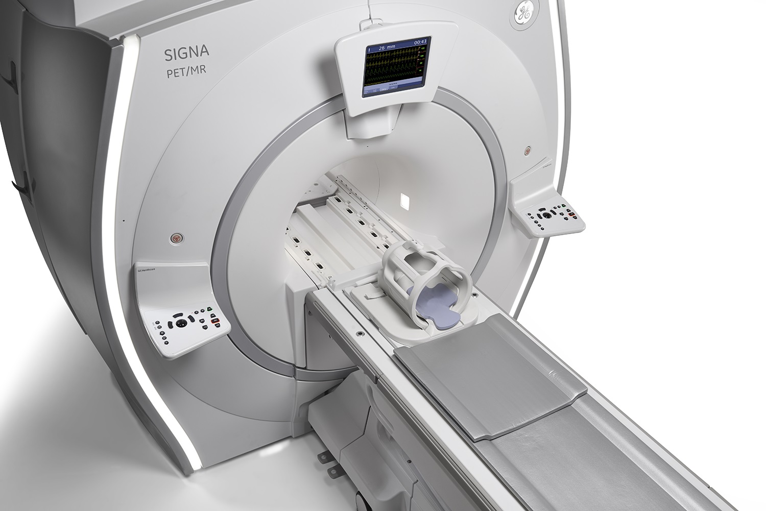
OHSU is the first hospital in the Pacific Northwest to acquire a PET/MRI hybrid scanner. This new state-of-the-art technology is located on the first floor of the Lamfrom Biomedical Research Building (LBRB) on OHSU's main campus. PET/MRI combines the excellent anatomic soft tissue detail of MRI with the molecular and functional information of PET.
How to order
-
Order panels are available in Epic for PET MRI. Please note, a “PET MRI” order alone does not include a full diagnostic MRI. To obtain a full diagnostic MRI in addition to the PET scan, two separate orders are required; a “PET MRI” order and a separate diagnostic “MRI” order. To make this easier, PET MRI order panels have been created for various clinical indications with the most common combinations selected by default and other options available for selection.
Our Diagnostic Radiology team will be happy to schedule patients through our scheduling line at (503) 494-8468. For questions or more information about PET/MRI, contact OHSU Nuclear Medicine and Molecular Imaging at petmri@ohsu.edu.
-
Please email us at petmri@ohsu.edu if you have questions or would like to request more information about PET/MRI.
-
Physicians and researchers interested in collaborating on new research projects can contact our Diagnostic Radiology Research team for more information at RADResearch@ohsu.edu.
Technical features
The GE SIGNA PET/MR features:
- Fully integrated system- 2 scans in one
- Simultaneous time of flight (TOF) PET imaging
- 3.0 T magnetic resonance imaging (MRI)
- 60 cm bore
- Weight limit- 227 kg (500 lbs)
Shared Research and Healthcare mission
In addition to the clinical benefits that this new technology brings, it enhances the collaboration between two OHSU missions, Research and Healthcare, amplifying our potential for developing new diagnostic and therapeutic tools for better patient outcomes. The Center for Radiochemistry Research is already working on new radiopharmaceuticals to complement the PET/MRI. In addition, this scanner offers the opportunity to the physicians and researchers of the Advanced Imaging Research Center, Knight Cancer Institute, Knight Cardiovascular Institute, and other OHSU research groups to collaborate on new clinical and research projects.
PET/MRI is a new hybrid imaging technology, combining two powerful diagnostic modalities, PET and MRI:
- PET or "Positron Emission Tomography" is an imaging modality that uses small amounts of radioactive materials called "radiotracers" to image the function of organs or systems within the body.
- MRI or "Magnetic Resonance Imaging," on the other hand, utilizes a strong magnetic field within the scanner and additional pulse sequences to image the organs and various structures in the body.
The hybrid and simultaneous imaging with PET/MRI combines the strengths of both modalities:
- Patient convenience is prioritized with the two studies performed at the same time.
- A more accurate diagnosis is possible, particularly if MRI is superior for the pathology in question.
- The ability to reduce the injected dose of radiotracers without compromising image quality due to the high sensitivity of the digital PET detectors.
Advantages of MRI
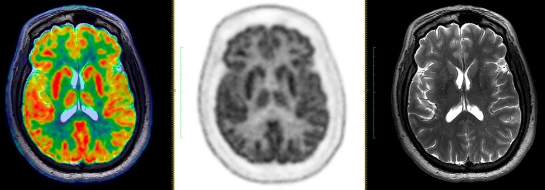
MRI provides superior soft-tissue contrast, helpful for multiple organs such as the brain, heart, liver, prostate, rectum, and reproductive tract. In addition, it allows the use of certain sequences, such as Diffusion-Weighted Imaging and MRI-specific contrast agents, that can add important information, particularly for oncology imaging.
Clinical applications
Particular scenarios for which PET/MRI might be the imaging modality of choice include:
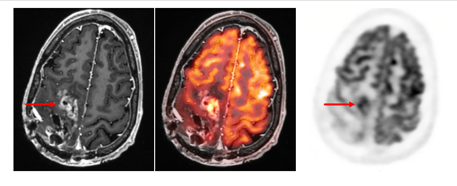
- Neurological conditions such as epilepsy, brain tumors, and dementia.
- Heart conditions such as sarcoidosis, myocarditis, and in the assessment of coronary atherosclerosis.
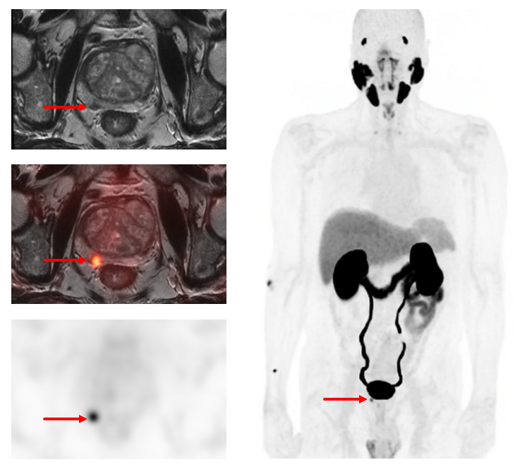
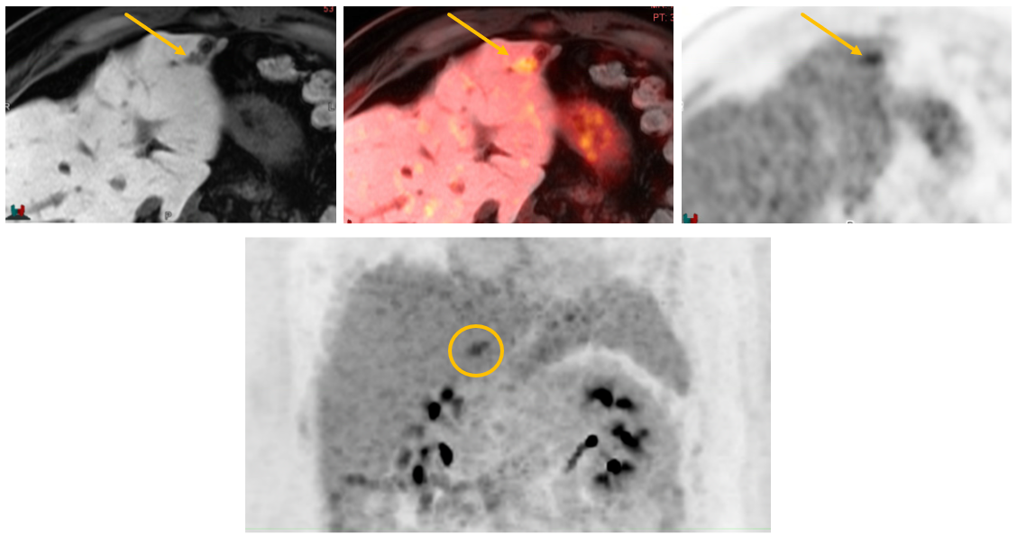
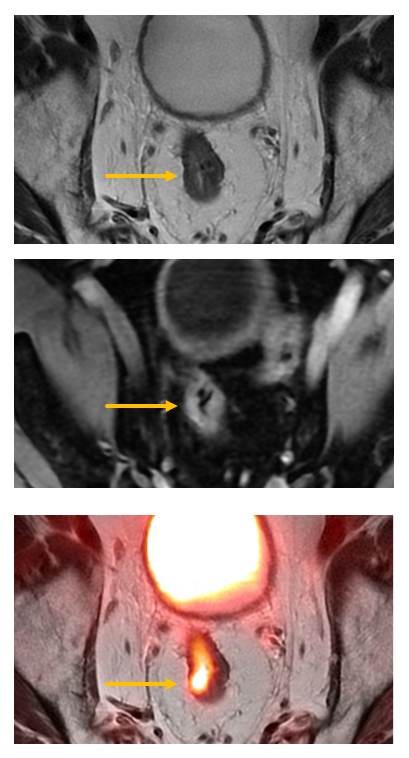
- Malignancies that are more optimally imaged with MRI, such as in cancers affecting soft-tissue areas of the body, including:
- Breast cancer
- Prostate cancer
- Rectal cancer
- Pelvic and gynecologic cancers
- Liver tumors
- Populations that would benefit from lower radiation doses.
Research
We are actively leveraging the creative talents of OHSU physicians & scientists with the new PET/MRI tools to make noninvasive discoveries about the human body. The combination of PET and MRI allows for an unparalleled understanding of disease processes through the ability to image cellular physiology while simultaneously capturing the highest quality anatomical images of tissues. In addition, we are continually adjusting old and developing new imaging techniques to create better views to understand the mechanisms that the human immune system uses to fight cancer. Using these new imaging approaches helps us improve disease diagnosis while also recognizing the importance of patient comfort by reducing imaging times to less than 10% of the current clinical standard.
The research applications of the PET/MRI system at OHSU are as varied as the human imagination, and our ultimate goal is to create new approaches to improve patients' lives. Researchers and physicians interested in collaborating on studies involving PET/MRI can contact an OHSU Radiologist to answer any questions. Likewise, patients interested in participating in imaging research at OHSU can talk to their doctor about opportunities or contact us via email at RadResearch@ohsu.edu.