Biomedical Imaging

Biomedical optics research image.

Optics is a branch of physics that examines the behavior and properties of light and the interaction of light with matter. Photonics is the science and technology of generating, controlling, and detecting photons, which are particles of light. The biophotonics lab conducts optics research with an emphasis on light propagation in biotissues.

The biophotonics resource center provides novel optical technologies for medical research and therapeutic and diagnostic tools for clinical medicine.

We research novel optical solutions to problems in basic science and clinical medicine.

Research projects

Image of spectral camera in process of imaging.

A spectral camera acquires x,y images at a series of wavelengths (λ). Then each x,y pixel becomes a reflectance spectrum, R(λ), which can be analyzed to specify tissue properties such as blood content and oxygen saturations, water content, light scattering, melanin content, etc.

We have two projects:

  1. The optics of appearance.
  2. Evaluating laser treatment of port wine stains in children.

The optics of appearance

We are acquiring spectral images of faces to specify the optical parameters (blood, hydration, melanin, scattering) that affect appearance.

Evaluating laser treatment of port-wine stains in children

Port-wine stains (PWS) are treated by a laser to thermally cause thrombus formation (clots), which yields tissue ischemia (lack of oxygen) and elicits a wound healing response that removes the PWS. But often treatment is incomplete. We are using a spectral camera to image the ischemia after laser treatment of PWS to find areas where the clot has loosened (reperfusion) which re-established blood flow and oxygenation such that treatment fails. The goal is to allow the doctor to find regions that need additional touch-up treatment before the patient leaves the clinic.

Optical fibers can deliver light to remote sites within the body. White light is delivered by one fiber and light transmitted/reflected from the tissue is collected by a 2nd fiber and routed to a spectrometer. The spectrum, R(λ) is analyzed to specify the blood content and oxygen saturation of the tissue.

Monitoring blood perfusion during laparoscopic surgery

Image of an optical fiber probe.

We are inserting an optical fiber probe through a trocar into the abdominal cavity during laparoscopic esophageal cancer surgery. The probe monitors the blood perfusion throughout the procedure and can give warning if the perfusion is held too low for too long, as blood vessels are tied off during the construction of a gastric conduit to replace the esophagus.

Figure showing data collected from using an optical fiber probe.

Interferometry can measure movements on the sub-nm scale. We use low-coherence light, achieved by a broad band of wavelengths, and lenses to restrict the interferometric measurement to a local region of interest.

Figure showing interferometry data from inner ear vibrations.

Vibrations of inner ear cochlear membrane

An optical coherence tomography (OCT) system is used to image the cochlear membrane of the inner ear, the ribbon of tissue down which sound waves propagate. The noise floor is 30 pm vibration, and in vivo vibrations are typical 0.1-10 nm. Figure: The vibrations of the cochlear membrane, a thin 100-µm-thick ribbon stretched between two bone supports. OC = organ of Corti. RM = Riessner's membrane.

Figure showing interferometry data from middle ear vibrations.

Clinical measurement of vibrations of middle ear structures

A similar OCT system is being developed as a hand-held otoscope to measure vibrations of the middle ear in the hearing clinic. Conductive hearing loss due to the ossicles (small ear bones) not properly moving in response to sound can be evaluated, without surgical violation of the typanic membrane. OCT can see through the typmanic membrane. Figure: TM = typmanic membrane. IN = incus. Ma = malleus.

Figure showing confocal reflectance bedside pathology imaging.

Bedside pathology for imaging skin cancer during Mohs surgery

We are striving to change how Mohs surgery is conducted, by placing a bedside confocal microscope and developing a rapid staining protocol so that an image equivalent to an H&E stained biopsy can be acquired within 5 min. The Mohs surgeon could stay with the patient and finish multiple excisions of skin cancer, rather than leaving the patient waiting for 30-45 min waiting for histopathology to be done. The staining procedure stains nuclei with one fluorophore (image #1) and cytoplasm with another fluorophore (image #2), and uses reflectance to image collagen fiber bundles (image #3). Then the 3 images are false-colored and combined to yield a single picture that mimics an H&E stained specimen, which is the type of image the Mohs surgeon routinely uses. Figure: Left (c) is false color image of basal cell carcinoma in human patient. Right (d) is H&E stained biopsy (Dan Gareau).

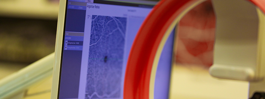
Study of immune cell trafficking in the iris of the the eye

Figure showing confocal reflectance imaging of mouse iris.

At right is an x-y image of a mouse eye in vivo at a particular depth z within the iris. The image shows dendritic immune cells that have acquired fluorophore-labeled antigen. Fluorescent cells are yellow. Gray image is backscatter of blue light by iris and other soft tissue structures.

Animated gif showing partially coherent fields imaging.

The propagation of light through complex structures, such as biological tissue, is a poorly understood phenomenon. Current practice typically ignores the coherence of the optical field. We use a novel Monte Carlo approach for propagating partially coherent fields through complicated deterministic optical systems. Random sources with arbitrary spatial coherence properties are generated using a Gaussian copula. Physical optics and Monte Carlo predictions of the first and second order statistics of the field are shown for coherent and partially coherent sources for a variety of imaging and non-imaging configurations. Excellent agreement between the physical optics and Monte Carlo predictions has been demonstrated in all cases.

We have developed a Monte Carlo-derived Green's function for the propagation of partially coherent fields. This Green's function, which is derived by sampling Huygens-Fresnel wavelets, can be used to propagate fields through an optical system and to compute first and second order field statistics directly. The concept is illustrated for a cylindrical f/1 imaging system. A Gaussian copula is used to synthesize realizations of a Gaussian-Schell model field in the pupil plane. The animated GIF shows the ensemble intensity for different cross-sections near the focus.

Ultimately, this formalism will be utilized to determine certain properties of a given optical system from measurements of the spatial coherence of the field at an output plane. Although our specific interests lie in biomedical imaging applications, it is expected that this work will find application to important radiometric problems as well.

Resources

Confocal laser scanning microscope, using both fluorescence and reflectance with 2-photon capability, for use with cells, small animals, and topical human skin sites

  • Optical coherence tomography, also for use with cells, small animals, and topical human skin sites
  • Optical micro angioGraphy (3D imaging of blood flow in microcirculation)
  • Polarized light camera for imaging superficial tissues over several cm2 field based on scattering by tissue structure
  • Spectral camera for imaging with spectroscopic contrast (blood, oxygen saturation, melanin, light scattering by tissue ultrastructure).
  • Optical spectroscopy via optical fibers (topical, via needles, via catheters)
  • Photodynamic therapy (targeted oxidative injury)
  • Biomechanical testing