The eye is known as the window to the body, and the cornea is known as the window to the eye. The clear outer layer at the front of the eye helps focus light, and as the outer-most structure of the eye, it’s vulnerable to damage.
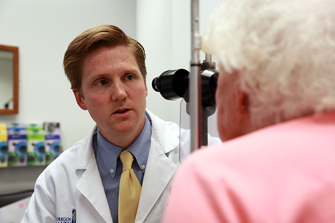
“The cornea is crucial to sight. Anything that traumatizes or damages the cornea can lead to very dramatic and rapid vision loss, because it's the first gateway through which light comes into the eye. When that initial gateway is closed, then it doesn't matter how well the other parts of the eye are working, the patient will have partial or total vision loss,” said Winston Chamberlain, M.D., Ph.D., Petti Professor of Ophthalmology at the Casey Eye Institute.
Elevating the art of cornea repair
Clinicians in Casey’s cornea division specialize in the diagnosis, treatment and management of a wide range of corneal conditions, from direct injuries to blinding genetic conditions. One of the most transformative treatments for corneal disease and abnormalities is corneal transplantation, a surgery that replaces a poorly functioning cornea with a donated cornea.
Corneal transplantation can save or restore sight for patients, such as those with inherited conditions or who experience problems that arise after cataract surgery. There are several current methods to choose from, but they are not equally effective. Some newer techniques require a high degree of expertise. Casey surgeons are refining these new methods and training other practitioners to use them.
“It's quite remarkable how much reconstruction we can do in this part of the eye versus other parts of the eye. We can restore most, if not all the function to the eye. That's one of the reasons I picked this subspecialty. We can get fantastic outcomes, and are often able to give patients their lives back,” said Chamberlain.
Dr. Chamberlain is a top corneal surgeon, performing more than 100 transplants a year, and serves as chair for the medical advisory board for the Eye Bank Association of America. He’s among the Casey faculty who are developing new, more effective surgical techniques that only require replacing one layer or section of the cornea, instead of the entire organ.
Thanks, in part, to clinical trials taking place at Casey, eye surgeons are now more likely to use a technique called single posterior layer transplant during corneal transplantation. Casey faculty are also pioneering a corneal transplant technique known as sutureless interlamellar keratoplasty (SILK) for addressing corneal ectasia, a condition that weakens the inner layers of the cornea.
The evolution of corneal transplantation
In a joint study with Stanford University and the University of San Francisco, called The Descemet Endothelial Thickness Comparison Trial (DETECT), Casey researchers compared the effectiveness of two distinct types of corneal transplantation and found that patients who received the thin layer transplant (referred to as DMEK) saw more significant improvement in their vision.
“In patients who had the thin layer transplant (DMEK), we saw improvements in vision even beyond what you can measure in a visual acuity chart – like their ability to see contrast – and they recovered faster. These thin layer transplants allowed them to get their vision back to normal at a faster rate, which is huge,” said Chamberlain, who was the co-author and co-designer of the study.
Retired OHSU operating room nurse Ginny McDaniel participated in the DETECT trial in 2016. She had been diagnosed with Fuchs dystrophy in both eyes and, had she not received the transplant, she would have eventually lost her vision. Today, McDaniel enjoys 20/20 vision and is grateful to her Casey clinicians, surgery team and cornea donor.
“I was on the liver transplant team at OHSU, and I understood the importance of donated organs. Little did I realize I was going to be the recipient of donated corneas. I'm very thankful for that.”
Building on success
Casey recently received funding from the National Eye Institute for a second DETECT study, bringing in a larger study population and adding a new element: testing the efficacy of Ripasudil eye drops, a drug developed in Japan that shows promise in restoring clarity to the cornea. Chamberlain’s hope is that the new drugs could help transplantation patients recover faster and allow the grafts to survive longer. The new study will start recruiting patients in the fall of 2022 and involve at least five clinical sites.
Better images, smarter diagnosis
Optical coherence tomography (OCT), pioneered by Casey’s associate director David Huang, M.D., Ph.D., has been a game changer for every field of ophthalmology, and the cornea is no exception. Creating precise images of a patient’s cornea is essential to diagnosing and treating corneal disease.
And now clinicians and researchers are pairing the power of OCT to make detailed tomographic and topographic maps of the cornea with artificial intelligence (AI), revolutionizing their ability to see problems early and guide decisions about how to approach treatment.
Using these tools, Yan Li, Ph.D., associate professor of ophthalmology, and colleagues are developing a protocol to image and track subtle changes in every layer of the cornea very early in corneal endothelial disease. OCT creates increasingly clear images of the cornea’s thickness and shape, and AI makes it possible to discern subtle abnormal patterns not visible to even the most skilled ophthalmologist. The combination significantly increases diagnostic accuracy.
“Clinical diagnosis can be subjective, and there can be biases between one doctor and another. Imaging data gives us objective facts,” said Dr. Li.
These new capabilities also make it possible to track patients with early signs of problems, and start treatment early with the least invasive options, like corneal collagen cross-linking, an outpatient procedure for the treatment of progressive keratoconus. What’s more, spotting issues early can often prevent the need for transplantation later on.
Human v. computer
Travis Redd, M.D., M.P.H, assistant professor of ophthalmology, recently published the results of a study in which his team taught a computer (using AI technology called convolutional neural networks, CNN) to use specific visual markers to distinguish between photographs of bacterial keratitis and fungal keratitis (aka corneal ulcers). These related conditions are notoriously hard to distinguish.
Corneal ulcers are caused by a fungal or bacterial infection in the cornea, often due to an injury to the eye or contact lenses. If left untreated, it can lead to blindness. The two types of infections require very different treatments.
Redd’s team asked study subjects to take a smart phone picture of their affected eye, and send it in for analysis. They sent the images to the computer lab and to a team of 12 cornea experts.
The CNNs were 81% accurate in diagnosing fungal ulcers and 75% accurate in diagnosing bacterial ulcers. Humans were better at pinpointing bacterial ulcers (88%), and significantly less accurate predicting fungal ulcers (56%).
“These findings could make a major impact on our ability to prevent blindness, globally,” said Redd.
Corneal damage is the 5th leading cause of blindness globally, and especially prevalent in less developed countries. As researchers and clinicians at Casey improve their ability to identify, heal and repair corneal damage, they are working towards stopping a significant percentage of preventable blindness here at home and, eventually, worldwide.