Voice & Swallowing - Diagnosis
During Your Visit - Diagnostic Studies
The larynx, situated deep in the neck, is pretty hard to see without special tools and diagnostic techniques. During your visit, your doctor may use one, or many, of these tools to figure out the correct diagnosis and treatment for your condition.
Indirect Laryngoscopy
The simplest form of laryngeal examination involves the placement of a small, angled mirror at the back of the throat. This allows the examiner to view the major structures of the larynx. Many of the smaller structures and their movements during normal speech, however, are difficult to see or to watch in motion. In patients with a strong gag reflex (easy to throw up), the usefulness of this technique may be limited. Still, this method allows a quick, easy, and painless gross examination of the larynx.
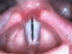
Flexible and Rigid Fiberoptic Laryngoscopy
These types of examination use fiberoptic (small cameras on cables) instruments to assist the examiner in looking at the larynx. The instruments possess a small camera in which the examiner can see the larynx. Usually, the picture seen with the instrument is fed via a camera lense into a digital recorder. This allows us to document your condition and also lets us keep a record for comparison with earlier, and later, exams.
In an examination with a flexible scope, you are given a spray to clear out your nose and to make it a little numb (anesthetize). The flexible fiberoptic scope is then sent through the nose and down the back of the throat into the pharynx. From this position, an image of the larynx and vocal folds (including their movement and position during breathing and speech) can be obtained and recorded.
An examination using a rigid scope involves placement of the tip of the instrument at the back of the tongue to provide a view of the throat. A prism at the tip of the scope allows the provider to see down into the larynx. This type of exam provides clear and highly magnified images of the vocal folds.
Videostroboscopy
During speech, the vocal folds vibrate far too quickly (over 100 times per second) to be examined with the naked eye. In order to overcome this, a strobe light is used to illuminate the vocal folds in slow-motion. A strobe emits bright pulses of light at evenly spaced intervals. If the frequency of those pulses is the same as the vibration frequency of the vocal folds, then the folds will appear "frozen" in time. If, however, the frequency of the strobe is slightly less than that of the vocal folds, the folds will appear to move in slow-motion.
This type of exam is important because it allows the vocal folds to be examined while you talk. Many problems with the larynx involve changes of this structure which in turn cause a change in the vibration of the vocal folds. During examination with a regular light source, these changes might go unnoticed. Stroboscopic examination allows the provider to see if any irregularities are present.
Electromyography (EMG)
This type of exam differs from those discussed above in that the larynx is not directly watched. Instead, how it is working is determined by certain changes which occur in the muscles of the larynx during contraction (tightening).
During electromyography, small needles are inserted through the skin into the muscles of the larynx. The activity of the muscles is then recorded while you are at rest (not speaking or swallowing) and during activity (speaking, breathing, and swallowing). The pattern of the activity gives the examiner clues to the nature of the disorder.
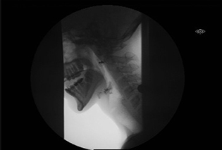
Videofluoroscopic Swallowing Study or a Modified Barium Swallow
Fluoroscopy is a technique, similar in principle to an ordinary X-ray. It is used to check and test how a patient is swallowing, if a swallowing problem is suspected. However, in contrast to the single picture of a simple x-ray, videofluoroscopy provides a continuous video image. This study is done with a radiologist and speech pathologist. In this exam, the patient swallows barium (a special mixture of chemicals) of different thickness. While the patient swallows, the examiner is able to see how the barium moves through the mouth and into the pharynx and esophagus.
Fiberoptic Endoscopic Evaluation of Swallowing (FEES)
As in Flexible Endoscopy, above, a FEES study uses a flexible scope to evaluate swallowing function. The nose is first decongested and anaesthetized (numbed) and then the scope is gently inserted through the nose in order to look at the structures of the larynx and pharynx. With the scope in place, the patient is then asked to do a variety of speech tasks which allow for the identification of any abnormalities in structure or function. After this, the patient receives something to swallow. In individuals who have a long history of aspiration and swallowing difficulties, this step to swallow may occur without food or liquid. In individuals who are currently eating and drinking, a variety of regular food and liquids are given to observe how the patient swallows. The examiner will assist if and when problems happen. During the examination, the patient and any family/caregivers in the room are able to see the examination in real-time on a monitor. This allows the examiner to teach students or residents, and to educate the patient, about the nature of the problem, evaluation of various swallowing strategies, and methods to teach swallowing maneuvers and exercises. This examination and the techniques then can form the basis of swallowing treatment.
How to Make an Appointment
Please contact our office regarding scheduling appointments. Prior to scheduling an appointment, our providers often need to review chart notes related to the reason you want to be seen in order to coordinate any additional tests they may need in order to make the most of your visit. In some cases, we will require a referral from your primary care physician. We would be happy to discuss this with you to ensure we have all we need to expedite the scheduling process. Please contact us by phone or email to begin the process.
Phone: NW Clinic for Voice & Swallowing 503-494-5947
Email: Otolaryngology -ENT Clinic
If you have a question for one of our providers, please email us.