Ultrasound and Fetal Monitoring
The OHSU Center for Women’s Health offers expert specialists and state-of-the-art technology to track your baby’s health and development. You’ll find:
- Advanced screening for birth defects and other prenatal conditions.
- Wireless monitoring of your baby’s heart rate and vital signs.
- Maternal Fetal Medicine specialists and radiologists who specialize in pregnancy care.
- Genetic counselors to help you understand your options.
- High-resolution 3D and 4D (with motion) ultrasound if medically needed.
Ultrasound monitoring
Ultrasound is an important tool for monitoring your pregnancy. We do scans throughout your pregnancy to check your baby’s growth and to catch any problems early. It is safe for both you and your baby.
Why have ultrasound?
During an ultrasound, we can see your baby as they move in your uterus. We measure your baby’s heart rate and growth, and look for signs of abnormalities. Ultrasounds also can help us determine:
- How long you’ve been pregnant
- Your due date
- The sex of your baby
- The number of babies and their positions
- Whether you have an ectopic pregnancy (in a fallopian tube)
- The position of the placenta and amount of amniotic fluid
- Your baby’s risk of genetic issues
How does ultrasound work?
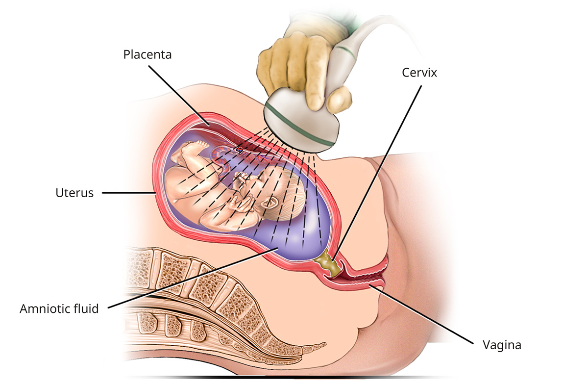
Ultrasound uses sound waves to create detailed images of your baby, umbilical cord and placenta. When the sound waves bounce off soft tissues, the echoes form an image, called a sonogram, on a computer screen.
It is done in two ways during pregnancy:
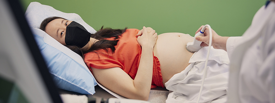
Transabdominal ultrasound: The technologist spreads gel on your belly and presses a probe on your skin to send sound waves into your uterus.
Ultrasound
Transvaginal ultrasound: A wand-shaped probe is inserted into your vagina for a closer look at your uterus from inside your body. This is usually done early in pregnancy or when we need to see your cervix better.
We also offer:
- Ultrasounds on the same day as your regular prenatal checkup in your provider’s office or in the nearby prenatal imaging center. An imaging specialist will talk with you about the results and answer your questions right away.
- Expert radiologists who specialize in diagnostic imaging for pregnancy using ultrasound and magnetic resonance imaging (MRI), when needed.
- Genetic counselors to address any concerns and to help you understand options for more screening and testing.
- Ultrasound screenings in your first trimester, at about 20 weeks and in your third trimester. If you have a high-risk pregnancy, you may have them more often.
- High-resolution 3D and 4D ultrasounds when medically needed. These provide clear views of your baby’s face, limbs and other body parts to help us diagnose physical issues, such as a cleft lip, and developmental conditions.
Ultrasound types and timing
-
How it’s done: Transvaginal ultrasound.
Why it’s done:
- To verify your pregnancy: An early ultrasound can confirm that your pregnancy is progressing normally.
- To determine your due date: We measure the size of your baby to calculate the expected due date. This is particularly important if:
- You have an irregular menstrual cycle.
- You aren’t sure when you had your last period.
- You conceived while breastfeeding or soon after stopping the pill.
- To see the number of babies: About 2% of women who conceive without fertility help have two or more babies. Fertility treatments increase odds to about 10%. An early ultrasound can show how multiple babies are developing and whether they share a placenta. Your care team will talk with you about adjusting your nutrition and care plan.
- To check the location of your pregnancy: Sometimes a fertilized egg implants in a fallopian tube instead of the uterus. This is called an ectopic pregnancy. It must be ended, because the fallopian tube cannot support an embryo. A tube rupture poses a serious health risk.
- To identify miscarriage risk: We look for problems in your uterus, placenta or embryo that might lead to a miscarriage.
- To check viability: If you have pain or bleeding, or if you have had a miscarriage or ectopic pregnancy, an early check can be reassuring or find any potential problem.
-
How it’s done: It’s usually a transabdominal ultrasound. Sometimes a transvaginal ultrasound is needed for a closer look at your baby.
Why it’s done:
- To measure nuchal translucency: Nuchal translucency is the clear space in the tissue of your developing baby's neck. We measure it to help assess your baby's risk of Down syndrome, other chromosomal abnormalities and major congenital heart problems.
- To check your baby’s anatomy: We look at your baby’s limbs, head, brain, spine, stomach and bladder to see if they appear to be forming normally. We also look to see if the umbilical cord is attached right.
- To confirm the number of babies.
- To confirm the baby’s gestational age and your due date.
- Next steps: If a potential issue is found, you can make a same-day appointment with our genetic counselors to discuss results. The nuchal translucency measurement alone cannot tell you the risk of Down syndrome or another condition. Other tests can help us confirm your baby’s condition or better calculate risk. Our genetic counselors can help you understand your options.
-
How it’s done: Transabdominal ultrasound.
Why it’s done:
- To check your baby’s anatomy: We examine each body part in detail. We pay special attention to the brain, face, spine, heart, stomach, bowel, kidneys and limbs.
- To determine sex (if desired): We can usually tell you if you are having a boy or a girl. In some cases, the genitals are not visible because your baby is facing away or has crossed legs. If you’d rather not know, you can ask us not to share results.
- To check your baby’s heart: In special cases, a fetal heart specialist may examine your baby’s heart and connecting blood vessels. This scan is recommended if you have a family history of heart issues or if we found increased nuchal translucency on a scan.
- For women at risk of early delivery: If you have a high risk of delivering your baby early, you may also have a transvaginal scan to check your cervix. Risk of early delivery rises with:
- A multiple pregnancy
- Previous early birth
- Abnormalities of the uterus
- Previous cervical surgery
-
How it’s done: Transabdominal ultrasound.
When it’s done: You may not need an ultrasound in your third trimester. If you or your care team has any concerns, you may have one scan or a series of scans to monitor your baby’s health and development.
Why it’s done:
- To check your baby’s growth and well-being: We measure your baby, estimate weight, and check movement.
- To check your baby’s position for birth: We look to see if your baby’s head is pointing down or up.
- To monitor possible complications: We do more scans if you have a condition such as preeclampsia or diabetes, or if your baby shows signs of not growing or moving as expected.
- To measure your amniotic fluid: Low amniotic fluid late in pregnancy can be a sign of problems with your placenta, a tear in the membrane, birth defects or other complications.
- To check on multiple babies.
Fetal monitoring
What is fetal monitoring?
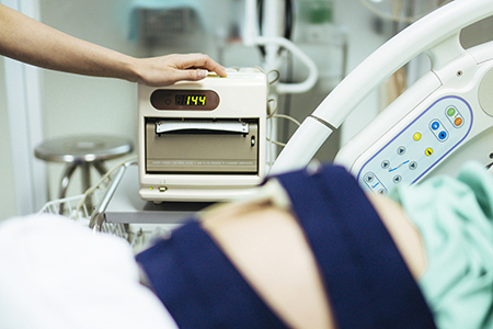
Before or during labor and childbirth, we can monitor your baby’s heartbeat and blood flow with electronic sensors on your belly. These monitors are safe. They give us important information about the well-being of your baby.
Why would I need fetal monitoring?
We may recommend fetal monitoring if you have:
- A high-risk pregnancy or medical condition, such as diabetes or preeclampsia.
- A history of complications with a previous pregnancy.
- A decrease in the movement of your baby.
- An injury or fall during pregnancy.
- An overdue pregnancy (41-42 weeks).
Nonstress test
What it is: This is the most common way we check your baby’s heart rate before birth. It’s called a “nonstress test” because it monitors your baby’s heart rate without stressing your baby. A healthy baby’s heart rate will rise and fall as it moves, without stimulation.
How it’s done: We strap monitors to your belly to check your baby’s heart rate while you rest. We do not give you any medication to cause your baby to move. There is no pain or risk to you or your baby.
Why it’s done: We may recommend this test if you or your providers have any concerns about your baby’s health or movement. Or you might have it if you have an injury or other complication.
Monitoring during labor and childbirth
Wireless fetal monitors track your baby’s heartbeat and your contractions during labor and childbirth. You may use them constantly or intermittently, depending on your preference, your baby’s condition and any complications. Talk with your care team about the best plan for monitoring your baby.
Related services
Advanced diagnostic imaging: We offer fetal MRI (magnetic resonance imaging) and fetal echocardiogram for more detailed views of your baby’s body, brain and heart.
Prenatal screening and genetics: Our genetic counselors can help you understand your risks for birth defects and genetic conditions. They can also discuss your testing options.
Fetal care: If your ultrasound finds signs of a birth defect or other condition, you may be referred to the Fetal Care Program at OHSU Doernbecher Children’s Hospital. Our expert team offers the most advanced care for you and your baby in the Northwest.
Learn more
- What is Ultrasound?
- Ultrasound Pregnancy, MedlinePlus, U.S. National Library of Medicine
- Ultrasound: Sonogram, American Pregnancy Association
- Fetal Monitoring, Journal of Midwifery and Women’s Health
For patients
Call 503-418-4200 to make an appointment.
Locations
Physicians Pavilion
3270 S.W. Pavilion Loop, Suite 420
Portland, OR 97239
Hillsboro Medical Center
8th Avenue Medical Plaza
364 S.E. 8th Ave., Suite 201
Hillsboro, OR 97213
Free parking for patients and visitors