Welcome!
The Vollum Institute is a privately endowed research institute at Oregon Health & Science University dedicated to basic research that will lead to new treatments for neurological and psychiatric diseases. Vollum scientists have broad-ranging interests that coalesce around molecular neurobiology and cellular physiology. Their work has transformed the field of neuroscience and, in particular, has provided important advances in the study of synaptic transmission, neuronal development, neurotransmitter transporters, ion channels and the neurobiology of disease.
Learn more about the Vollum's mission
Vollum Seminar Series
The Vollum seminar series is on hiatus and will return in the fall. Please check back for more information in late September.
Friday Work–In–Progress Talks
The Friday "work-in-progress" (WIP) seminars occur weekly during the academic year and provide an opportunity for postdoctoral fellows and graduate students to share their current research projects in an interactive and less formal environment.
The WIP seminar series is on hiatus and will return in the fall. Please check back for more information in late September.
Research highlights
A highly sensitive genetically encoded red cAMP sensor for multiplex imaging in vivo.
Wang L, Li L, Li X, Guo L, Luo B, Han Y, Fang S, Wang S, Shao R, Chen J, Stehle D, Zhang J, Mu Y, Zhu C, Du Y, Zhong H, Li Y, Chu J. Nat Commun. 2026 May 25. doi: 10.1038/s41467-026-73138-5. PMID: 42185274.
Grin2a knockout in adolescent dopamine neurons disrupts prediction error signaling and produces a phenotype relevant to schizophrenia.
Kielhold ML, Jacobs DS, Torrado Pacheco A, Lefner MJ, Bogachuk AP, Lebowitz JJ, Langdon AJ, Williams JT, Zweifel LS, Moghaddam B. Mol Psychiatry. 2026 May 13. doi: 10.1038/s41380-026-03632-1. PMID: 42129480.
Stochastic growth and ligand-receptor interaction-mediated stabilization generate stereotyped dendritic arbors.
Shi R, Ho XY, Tao L, Bayless-Edwards L, Taylor CA, Zhao T, Zou W, Lizzappi M, Eichel K, Mao T, Shen K. Nat Neurosci. 2026 May 4. doi: 10.1038/s41593-026-02278-0. PMID: 42082815.
Comprehensive 3D mapping reveals distinct spatial gradients of genetically-identified SST, PV, and TH interneurons across the mouse caudoputamen.
Muhsinov JM, Iliakis EA, Tu W, Ramirez AN, Muniak MA, Wasilczuk AZ, Davatolhagh MF, Proekt A, Mao T, Fuccillo MV. Front Cell Neurosci. 2026 Apr 16;20:1795921. doi: 10.3389/fncel.2026.1795921. eCollection 2026.PMID: 42078083.
A discrete serotonergic circuit involved in the generation of tinnitus behavior.
Yu MT, Dai ZY, Wang SX, Wang KJ, Qin CH, Zhou YM, Guo XT, Pan CC, Sun JQ, Sun JW, Chen WH, Jin Y, Wu QW, Trussell LO, Tang ZQ. Proc Natl Acad Sci U S A. 2026 Apr 28;123(17):e2509692123. doi: 10.1073/pnas.2509692123. Epub 2026 Apr 20. PMID: 42008671.
PTEN regulates starburst amacrine cell dendrite morphology during development.
Bracha TW, Luong N, Leffler J, Sivyer B, Wright KM. Development. 2026 May 1;153(9):dev204980. doi: 10.1242/dev.204980. Epub 2026 May 7. PMID: 41968902.
Flexible ensheathment of axons enables myelination of complex CNS networks.
Call CL, Neely SA, Early JJ, James OG, Zoupi L, Williams AC, Xu YKT, Chandran S, Lyons DA, Monk KR, Bergles DE. Nature. 2026 Apr 1. doi: 10.1038/s41586-026-10312-1. PMID: 41922759.
Tetrazine Functionalized Graphene Enables Capture of Ultra-Low Concentrations of Biomacromolecules.
Gupta RK, Jeong H, Goehring A, Gouaux E, He M. Small. 2026 May;22(28):e13723. doi: 10.1002/smll.202513723. Epub 2026 Mar 25. PMID: 41880224.
Lysosomal down-regulation of the mu opioid receptor is opposed by the Retromer complex.
Dagunts A, Adoff H, Novy B, Ameur LB, De Maria M, Saunders A, Lobingier BT. Sci Adv. 2026 Mar 20;12(12):eadx8715. doi: 10.1126/sciadv.adx8715. Epub 2026 Mar 20. PMID: 41861010.
The molecular basis of force selectivity by PIEZO2.
Mulhall EM, Yarishkin O, Hill RZ, Koster AK, Patapoutian A. Nature. 2026 May;653(8113):297-305. doi: 10.1038/s41586-026-10182-7. Epub 2026 Mar 4.PMID: 41781615.
The protocadherin-15-LHFPL5 tip link complex is a heterotetrameric assembly in hair cell stereocilia.
Clark S, Mitra J, Elferich J, Goehring A, Ge J, Ha T, Gouaux E. Biophys J. 2026 Feb 10:S0006-3495(26)00092-5. doi: 10.1016/j.bpj.2026.02.003. Online ahead of print. PMID: 41668373.
Molecular assemblies and pharmacology of cerebellar GABAA receptors.
Sun C, Jahncke JN, Wright KM, Gouaux E. Proc Natl Acad Sci U S A. 2026 Feb 10;123(6):e2524504123. doi: 10.1073/pnas.2524504123. Epub 2026 Feb 6. PMID: 41650215.
Hypoxia-inducible factor 1 protects neurons from Sarm1-mediated neurodegeneration.
Meraner P, Avetisyan A, Swift K, Cheng YC, Barria R, Freeman MR. Cell Rep. 2026 Feb 24;45(2):116873. doi: 10.1016/j.celrep.2025.116873. Epub 2026 Jan 22. PMID: 41575852.
Cryo-EM of autoantibody-bound NMDA receptors reveals antigenic hotspots in an active immunization model of anti-NMDAR encephalitis.
Kim J, Jalali-Yazdi F, Jones BE, Westbrook GL, Gouaux E. Sci Adv. 2026 Jan 16;12(3):eaeb4249. doi: 10.1126/sciadv.aeb4249. Epub 2026 Jan 14. PMID: 41533802.
Reversible lipid-mediated pH-gating of connexin-46/50 by cryo-EM.
Jarodsky JM, Myers JB, Reichow SL. Nat Commun. 2026 Jan 12;17(1):1606. doi: 10.1038/s41467-026-68311-9. PMID: 41526355.
Renal PIEZO2 is an essential regulator of renin.
Hill RZ, Nelson JW, Gyarmati G, Medrano S, Shirvan S, McCormick JA, Burquez S, Ahmed J, Eng DG, Wysocki J, Dubin AE, Servin-Vences MR, Lakshmanan A, Gomez RA, Sequeira-Lopez MLS, Shankland SJ, Batlle D, Miner JH, Peti-Peterdi J, Patapoutian A. Cell. 2025 Dec 4:S0092-8674(25)01309-1. doi: 10.1016/j.cell.2025.11.013. PMID: 41349545.
Astrocyte gap junctions and K ir channels contribute to K+ buffering and regulate neuronal excitability.
Bojovic D, Dagostin A, Sullivan SJ, Emery B, von Gersdorff H, Mishra A. Front Cell Neurosci. 2025 Nov 20;19:1571218. doi: 10.3389/fncel.2025.1571218. eCollection 2025. PMID: 41356497.
The photoswitchable cannabinoid azo-HU308 enables optical control of Ca2+ dynamics in INS-1 β-cells via off-target effects on TRPC channels.
Viray AEG, Frank JA. FEBS Open Bio. 2025 Nov 2. doi: 10.1002/2211-5463.70146. PMID: 41178137.
Targeting Tmem63b and Piezo2 in C-fiber low-threshold mechanoreceptors: Limitation of Vglut3-IRES-Cre.
Orlin DJ, Muñoz A, Berryman S, Semidey D, Murthy SE. Biophys J. 2025 Nov 1:S0006-3495(25)00733-7. doi: 10.1016/j.bpj.2025.10.043. Online ahead of print. PMID: 41176617.
Altered Primary Somatosensory Neuron Development in a Pten Heterozygous Model for Autism Spectrum Disorder.
Fernandez A, Sarn N, Eng C, Wright KM. Autism Res. 2025 Sep 12. doi: 10.1002/aur.70119. Online ahead of print. PMID: 40940651.
Human brain cell types shape host-rabies virus transcriptional interactions revealing a preexisting pro-viral astrocyte subpopulation.
Feige L, Young K, Cerapio JP, Kozaki T, Kergoat L, Libri V, Ginhoux F, Hasan M, Ben Ameur L, Chin G, Goode Z, Bourhy H, Saunders A. Cell Rep. 2025 Aug 25;44(9):116159. doi: 10.1016/j.celrep.2025.116159. Online ahead of print. PMID: 40864551.
FBXW7 regulates MYRF levels to control myelin capacity and homeostasis in the adult central nervous system.
Collins HY, Doan RA, Li J, Early JE, Madden ME, Simkins T, Lyons DA, Monk KR, Emery B. Nat Commun. 2025 Aug 21;16(1):7822. doi: 10.1038/s41467-025-62715-9. PMID: 40841354.
TMEM63A, associated with hypomyelinating leukodystrophies, is an evolutionarily conserved regulator of myelination.
Halford J, Senatore AJ, Berryman S, Muñoz A, Semidey D, Doan RA, Coombs AM, Noimany B, Emberley K, Emery B, Monk KR, Murthy SE. Proc Natl Acad Sci U S A. 2025 Jul 29;122(30):e2507354122. doi: 10.1073/pnas.2507354122. Epub 2025 Jul 21. PMID: 40694323.
A high-performance genetically encoded sensor for cellular imaging of PKC activity in vivo.
Yahiro T, Bayless-Edwards L, Jones JA, Zhuo Y, Ma L, Qin M, Mao T, Zhong H. Nat Commun. 2025 Jul 10;16(1):6378. doi: 10.1038/s41467-025-61729-7. PMID: 40640216.
New images map key membrane protein in brain related to stroke
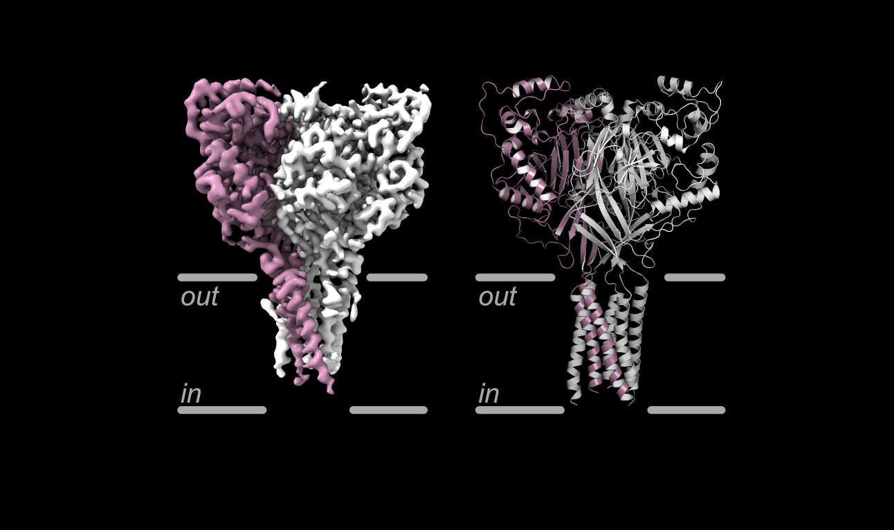
Scientists have for the first time mapped in exquisite, three-dimensional detail six major conformations of a membrane in the brain related to learning, memory and fear-related behavior.
The study, led by researchers at Oregon Health & Science University, published today in Nature Structural & Molecular Biology.
Researchers used state-of-the-art cryo-electron microscopy housed in OHSU’s South Waterfront Campus to capture the most detailed view yet of a specific type of membrane protein: an acid-sensing ion channel known as ASIC1a.
The findings could form the blueprint for drug development useful in treating stroke, said senior author Isabelle Baconguis, Ph.D., assistant professor in the OHSU Vollum Institute.
OHSU researchers and collaborators receive PMedIC Innovation awards
The annual Precision Medicine Innovation Co-Laboratory Innovation Grant supports collaborative pilot projects annually between OHSU and Pacific Northwest National Laboratory researchers as part of the formal partnership called PMedIC.
For fiscal year 2027, two grants were awarded to OHSU and PNNL research teams:
James Frank, Ph.D., associate professor of chemical physiology and biochemistry in the OHSU School of Medicine with a joint appointment at the OHSU Vollum Institute, and Vivian Lin, Ph.D., chemist with PNNL: “Development of Bifunctional Phytocannabinoid Probes for Proteome-Wide Target Identification.”
Cody Call research published in Nature
Cody Call, Ph.D., researcher at OHSU’s Vollum Institute, is first author of a study recently published in the journal Nature.
Call performed zebrafish experiments and electron microscopy analysis while in the lab of Kelly Monk, Ph.D., who is second to last author of the study, “Flexible ensheathment of axons enables myelination of complex CNS networks.”
Many nerve fibers in the brain are wrapped in a fatty insulation called myelin, which allows electrical signals to travel quickly and is essential for healthy brain circuit function. The cells that produce myelin, called oligodendrocytes, each grow multiple arm-like processes. Scientists long believed that each arm wrapped exactly one segment of a nerve fiber in a uniform, synchronized way: like tape around a single stretch of wire. But this model did not explain how the brain insulates complex, highly branched networks, where physical obstacles often get in the way. This paper challenges that idea.
Dr Marc Freeman elected to the American Academy of Arts and Sciences
Marc Freeman, Ph.D., director and senior scientist of the OHSU Vollum Institute, has been elected to the American Academy of Arts and Sciences. The Academy, whose members include Albert Einstein, Martin Luther King, Jr., and Maria Mitchell, is both an honorary society that recognizes and celebrates the excellence of its members and an independent research center convening leaders from across disciplines, professions and perspectives to address significant challenges. Freeman is one of more than 250 members who will be inducted in a ceremony in October 2026.
Research links tinnitus with serotonin
Researchers with Oregon Health & Science University and Anhui University in China found in a mouse model that elevated levels of the neurotransmitter serotonin in the brain also resulted in elevated behavioral symptoms of tinnitus.
The findings should be especially meaningful for millions of people around the world who suffer from tinnitus, said co-senior author Laurence Trussell, Ph.D., professor of otolaryngology in the OHSU School of Medicine and a scientist in the OHSU Vollum Institute and Oregon Hearing Research Center.
Read the full article
Listen or read OPB Interview
Trussell Lab
Once Thought To Support Neurons, Astrocytes Turn Out To Be in Charge
The human brain is a vast network of billions of neurons. By exchanging signals to depress or excite each other, they generate patterns that ripple across the brain up to 1,000 times per second. For more than a century, that dizzyingly complex neuronal code was thought to be the sole arbiter of perception, thought, emotion, and behavior, as well as related health conditions. If you wanted to understand the brain, you turned to the study of neurons: neuroscience.
But a recent body of work from several labs, published as a trio of papers in Science in 2025, provides the strongest evidence yet that a narrow focus on neurons is woefully insufficient for understanding how the brain works. The experiments, in mice, zebra fish, and fruit flies, reveal that the large brain cells called astrocytes serve as supervisors. Once viewed as mere support cells for neurons, astrocytes are now thought to help tune brain circuits and thereby control overall brain state or mood — say, our level of alertness, anxiousness, or apathy.
Scientists identify target to treat devastating brain disease
Scientists have identified a promising target for treatment of a devastating autoimmune disease affecting the brain.
The discovery could lead to the development of new therapies for a disease triggered by an attack on one of the key neurotransmitter receptors in the brain, the NMDA receptor. It also raises the potential for a blood test to detect a signal of the condition and enable earlier treatment with existing therapies.
The study from Oregon Health & Science University published today in the journal Science Advances.
Researchers identify kidney sensor that helps control fluid balance
A new study has identified a critical “pressure sensor” inside the kidney that helps the body control blood pressure and fluid levels. The finding helps explain how the kidneys sense changes in blood volume — something scientists for decades have known occurs but didn’t have a mechanistic explanation.
More news and accolades
- Dr Marc Freeman elected to the American Academy of Arts and Sciences
- Saunders lab's Cell Reports article describes the unique role of astrocyte innate immune signaling in combating viral infection
- Marissa Co named Simons Foundation Fellow
- Two graduate students named 2025 Lacroute Fellows
- Vollum neuroscientist achieves rare distinction, earns $5.4 million award
Recognition for our early career awardees
Graduate students and postdoctoral fellows are usually supported by research grants to individual faculty or by institutional training grants from the NIH. However, a sought-after perk for trainees is to obtain an individual fellowship from federal sources or foundations. Such awards are an honor and also provide important financial support for the trainee and their lab. Graduate students and postdoctoral fellows in the Vollum Institute have been remarkably successful in obtaining these awards over the past few years. This is a credit to the quality of the trainees and the support they receive from their mentors. Congratulations to all.
-
Two predoctoral students in the Neuroscience Graduate Program at OHSU were recently awarded the 2025 Lacroute Fellowship. This fellowship is made possible through the philanthropic support of a generous donor.
The Lacroute Fellows Program supports exceptional graduate students conducting innovative research in the Vollum Institute/OHSU Neuroscience Graduate Program. The one-year fellowships cover $24,000 of the students’ stipend and provide a $1,000 allowance for related academic expenses, such as attending scientific conferences or courses.
Congratulations to the 2025 fellows:
- Hoa Trinh, Freeman Lab
- Jed Syrenne, Murthy Lab and Reichow Lab
2024 Lacroute Fellows
2023 Lacroute Fellows
2022 Lacroute Fellows -
- Cody Call, Ph.D., Monk Lab
NINDS K99: “A novel role for cell cycle regulators in oligodendrogenesis and myelination.” - Marissa Co, Ph.D., Wright lab
Simons Foundation Fellow - Armani Del Franco, Ph.D., Monk Lab
Natl Multiple Sclerosis Society: "In-vivo investigations of oligodendrocyte-astrocyte interactions responsible for myelin integrity." - Nathaniel Ghena, Ph.D., Freeman Lab
NINDS F32: “Exploring role of astrocytic adhesion GPCR Remoulade in neuronal remodeling.” - Dongeun Heo, Ph.D., Monk/Freeman Labs
Collins Medical Trust: "Novel role of SARM1 in the regulation of oligodendrocyte development and myelination." - James Jones, Ph.D., Mao Lab
NIMH F32: "Intracellular signaling dynamic control of synaptic responses in the basal ganglia." - Damien Rasmussen, Ph.D., Gouaux Lab
NIH Natl Inst of Mental Hlth: “Conformational mechanisms underlying allosteric regulation of the human serotonin transporter.” - Colin Wakeham, Ph.D., von Gersdorff Lab
NIH Natl Eye Inst: "Characterizing the Unique Biophysical Properties of the CBC2 OFF Cone."
- Cody Call, Ph.D., Monk Lab
-
- Madison Hupp, Freeman Lab
NINDS F31: "Neuronal and glial mechanisms regulating Pair1 local pruning." - Arielle Isakharov, Wright Lab
NEI F31 Predoctoral Fellowship: "Genetic analysis of the Robo3+ glycinergic amacrine cell." - Chloé Le Moing, Jackman Lab
NINDS F31: "Examining the role of synaptic facilitation in cortical network function and behavioral flexibility." - Kayla Maanum, Mao Lab
NINDS F31: "Subcellular and mesoscale circuit organization for taste and visceral processing." - Tony Muñoz, Murthy Lab
HHMI Gilliam Fellow: "Investigating Mechanosensors Involved in Swallowing Physiology." - Yessica Santana Agreda, Wright Lab
HHMI Gilliam Fellow: "Transcriptional Control of Starburst Amacrine Cell Specification and Maturation." - Erin Santos, Freeman Lab
HHMI Gilliam Fellow: "CamKII-Dependent Mechanisms of Astrocyte Ca2+ Signaling." - Frederika Sullivan, Wright Lab
National Science Foundation, Graduate Research Fellowship - Jed Syrenne, Murthy Lab and Reichow Lab
2025 Lacroute Fellow - Hoa Trinh, Freeman Lab
2025 Lacroute Fellow
- Madison Hupp, Freeman Lab
-
Congratulations to all of our graduate researchers in the Vollum/OHSU Neuroscience Graduate Program who received ARCS Foundation Scholar Awards from the ARCS Oregon Chapter!
First Year (2025–2028): Sandra Christina Salas
Second Year (2024-2027): Juliana Cuartas & Alexa Gonzalez
Third Year (2023-2026): Kayla Maanum, Melissa (Arisa) Sek & Frederika SullivanARCS Foundation Oregon is honored to present awards to these outstanding scholars chosen by the scholar selection committees at Oregon Health & Science University, Oregon State University and the University of Oregon. The ARCS Foundation Scholar Award is $18,000, payable over three years at $6,000 per year.
Learn more about these scholars and the ARCS Foundation Oregon -
Congratulations to the Neuroscience Graduate Program researchers who received 2025-2026 N.L. Tartar Trust Fellowships.:
James Jones (Mao Lab)
Milana Krush (Westbrook Lab)
Nina Luong (Wright Lab)
Joselinne Medrano (Williams Lab)
Yizhou Zhuo (Zhong Lab)The $2,000 grants are awarded annually by the OHSU School of Medicine as a means to support research endeavors and career development. Keep up the great work!