MRI Pelvis Sports Hernia WO MSK Protocol
Scan Notes:
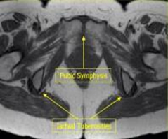
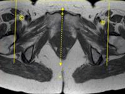
Center at symphysis pubis
If hardware is present:
- Do Axial and Coronal STIR instead of fat-sat mid-TE
- If with Contrast, do non-fat-sat T1 post-contrast
Last updated: 4/8/19
Charge as: Pelvis WO
Scanner preference: 3T or Ingenia only
Coil: Torso Coil
| Plane | Weighting | Mode | Slice | Gap | FAT SAT | FOV | Notes |
|---|---|---|---|---|---|---|---|
| AXIAL | T2 SPAIR | TSE | 3-4 mm | 0.5-1mm | SPAIR | both hips | 1 cm above acetabulum thru symphysis pubis |
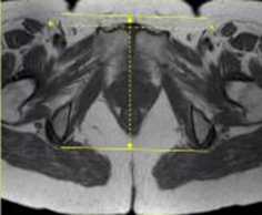
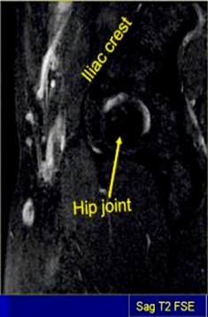
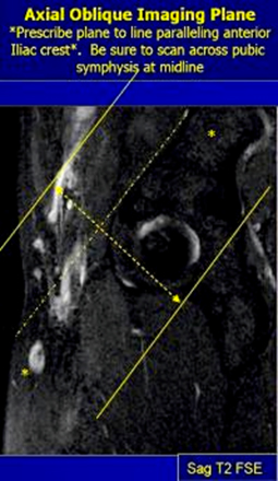
| AX OBL | PD (TE=20msec) | TSE | 3-4 mm | 0.5-1mm | None | 20cm | Parallel anterior iliac crest, from crest thru hip joint |
| AX OBL | T2 SPAIR | TSE | 3-4 mm | 0.5-1mm | SPAIR | 20cm | Parallel anterior iliac crest, from crest thru hip joint |
| SAG | T2 SPAIR | TSE | 3-4 mm | 0.5-1mm | SPAIR | 20cm | Ischial tuberosity to ischial tuberosity (NOT skin to skin) |
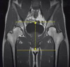
| COR | T1 | TSE | 3-4 mm | 0.5-1mm | None | both hips | 1cm anterior to symphysis pubis thru both ischial tuberosities |
| COR | T2 STIR (TE=50-60) | TSE | 3-4 mm | 0.5-1mm | STIR | both hips | 1cm anterior to symphysis pubis thru both ischial tuberosities |