MR Pituitary and Brain WWO Neuro Protocol
Last updated: 01/20/22
Charge as: Brain WWO
Scanner preference: Siemens 3T > Philips 3T > Siemens 1.5T
Coil: Head
| Plane | Weighting | Mode | Slice (mm) | Gap (mm) | FAT SAT | FOV (cm) | MPR | Notes |
|---|---|---|---|---|---|---|---|---|
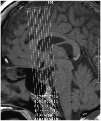
| SAG | T1 | 3D TSE | 0.7 - 1.0 | 0 | no | 23 | AXIAL, COR | Whole brain coverage. Cover at least 1cm above vertex through skull base. Cover entire nose. |
| AXIAL | T2 | 3D TSE | 1 | 0 | no | 23 | no | Whole brain coverage. Angle to corpus. Cover at least 1 cm above vertex through skull base. Cover entire nose. |
| AXIAL | DWI | EPI | 3 | 0.3 | no | 23 | no | Angle to Corpus. Cover Skull Base to Vertex. Send only B1000 & ADC. |
| AXIAL | SWI | 3D GRE | 2 | -1 | no | 23 | no | Angle to Corpus. Cover skull base to vertex. Ok to add slices. |
| COR | T2 | 2D TSE | 1.5 | 0 | no | 13 | no | Pituitary only. Angle perpendicular to corpus. |
Contrast injection
Power inject contrast @ 2cc/sec
Inject when the 1st dynamic ends and the 2nd dynamic begins
20cc saline flush @ 2.0 cc/sec
You may increase # of slices, but ensure that EACH DYNAMIC does NOT EXCEED 20 sec.
| Plane | Weighting | Mode | Slice (mm) | Gap (mm) | FAT SAT | FOV (cm) | MPR | Notes |
|---|---|---|---|---|---|---|---|---|
| COR | T1 | TSE DCE (dynamic) | 2 | 0.2 | no | 15 | no | Dynamic scans to cover the Pituitary only. Each Dyanamic should not exceed 20 seconds. |
| SAG | T1 | 3D TSE | 0.7-1.0 | 0 | no | 18 | AXIAL, COR | Small FOV centered on pituitary. L-R coverage 8 cm. Angle to interhemispheric fissure. |
| SAG | T2 FLAIR | 3D IR-TSE | 1 | 0 | YES | 23 | no | Whole brain coverage. Angle to interhemispheric fissure. Cover ears and nose. Spacing and gap are variable. |
| AXIAL | T1 | 3D TFE | 1 | 0 | no | 23 | no | Whole brain coverage. Angle to corpus. Cover at least 1cm above vertex through skull base. Cover entire nose. |