MR Pediatric Hip Perthes WWO Protocol
Scan Notes: It is absolutely critical that the pre-contrast and post-contrast images line up exactly so that there is no mis-registration on the subtracted images.
To this end: 1) immobilize the affected limb with a sandbag across the ankle; and 2) minimize patient stimulation during the administration of contrast by either using the power injector at a low flow rate (1ml/sec) or hand inject through an extension. Do not remove the patient from the scanner to facilitate the injection.
See the following references for more information: www.perthesdisease.org
Last updated:3/28/19
Charge as: Hips WWO
Scanner preference: 3T Ingenia ONLY - DCH7 or MR4
Coil: Torso or Cardiac
Scan Notes: Images must be checked by radiologist or radiology resident before giving contrast and before the patient gets off the table.
| Plane | Weighting | Mode | Slice | Gap | FAT SAT | FOV | Notes |
|---|---|---|---|---|---|---|---|
| COR | T1 | TSE | 4mm | 0mm | None | 24cm | |
| COR | T2 Fat Sat | TSE | 4mm | 0mm | SPAIR | 24cm | |
| AXIAL | T2 | TSE | 4mm | 0mm | None | 24cm | |
| COR | T1 Fat Sat | TSE | 4mm | 0mm | SPIR | 24cm | |
| SAG | T1 Fat Sat | TSE | 4mm | 0mm | SPIR | 18cm |
Contrast Injection
Use power injector at reduced flow rate (1ml/sec) or extension tubing. Do not remove patient from scanner to perform injection. Wait 2 minutes after injection is complete before starting post-contrast scans.
| Plane | Weighting | Mode | Slice | Gap | FAT SAT | FOV | Notes |
|---|---|---|---|---|---|---|---|
| COR | T1 Fat Sat | TSE | 4mm | 0mm | SPIR | 24cm | Perform manual subtraction (post-pre) |
| SAG | T1 Fat Sat | TSE | 4mm | 0mm | SPIR | 18cm | Perform manual subtraction (post-pre) |
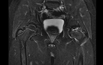
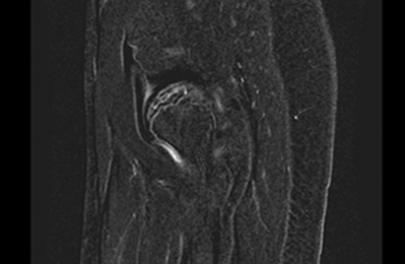
Post-Processing: Perform manual subtractions of the coronal and sagittal T1 Fat Sat sequences – postcontrast – precontrast = subtracted image. It should look like this: