MR Osteochondral Planning Knee WO Protocol
Also known as Episurf MRI Protocol Knee Endoscopy.
Dr. Crawford must be indicated as Ordering or Attending physician.
- Run as built in
- Don't change parameters
- Don't change FOV
- Ok to add slices
Last updated: 9/27/19
Charge as: Knee WO
Scanner preference: MR3 or MR4 only
Coil: Knee Coil
-
POSITIONING
- Place the knee and coil as close to isocenter as possible.
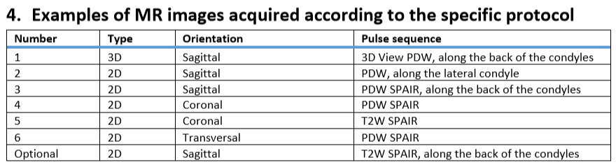
3D SEQUENCE
- Do not change the FOV or the matrix size. 3D representations of the knee are created from this sequence.
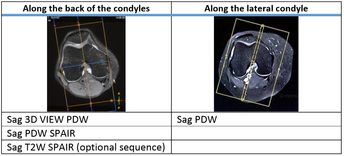
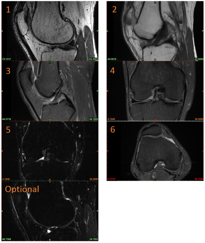
SAGITTAL SEQUENCES
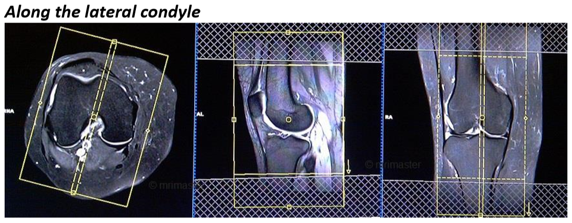
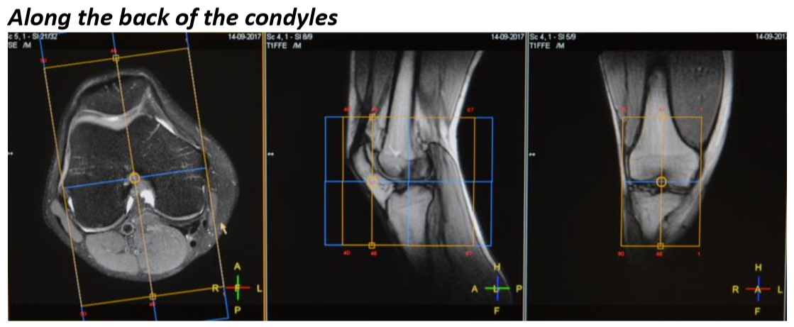
- Not all SAG sequences are oriented the same. Please follow scan guidelines for all sequences.
FIELD OF VIEW
- FOV for all sequences will cover the entire femoral bone and articulating cartilage.
SAG T2 SPAIR
- Run the SAG T2 SPAIR if the patient has a history of prior meniscal surgery.
USE OF FOLDOVER SUPPRESSION AND SENSE
- Use fold-over suppression if artifacts are interfering with the bones or articulating cartilage.
- OK to use SENSE as long as superior image quality is maintained.
| Plane | Weighting | Mode | Slice | Gap | FAT SAT | FOV | Notes |
|---|---|---|---|---|---|---|---|
| SAG 3D VIEW PDW (PHILIPS) | 3D | NONE | Do not change | Angle perpendicular to the back of the condyles | |||
| SAG 3D SPACE (SIEMENS) | 3D | NONE | Do not change | Angle perpendicular to the back of the condyles | |||
| AXIAL | PD SPAIR | 2D | 3 | 0.3 | SPAIR | Do not change | |
| SAG - Angle parallel to the lateral condyle | PD | 2D | 3 | 0.3 | NONE | Do not change | Angle parallel to the lateral condyle |
| SAG | PD SPAIR | 2D | 3 | 0.3 | SPAIR | Do not change | Angle perpendicular to the back of the condyles |
| COR | PD SPAIR | 2D | 3 | 0.3 | SPAIR | Do not change | |
| COR | T2 SPAIR | 2D | 3 | 0.3 | SPAIR | Do not change | |
| SAG (Optional - run if hx prior meniscal surgery) | T2 SPAIR | 2D | 3 | 0.3 | SPAIR | Do not change | Angle perpendicular to the back of the condyles. Run if the patient has previously undergone a meniscal surgery. |