MR MS Brain WWO Neuro Protocol
Last updated: 10/18/2023
Charge as: Brain WWO
Scanner preference: 3T only
Coil: Head
New Neuroquant Workflow Changes as of 10/18/2023
See instructions below for changes to the workflow for Neuroquant images
-
VIP Notes for MRI Tech
Exam data must include: Age/DOB, Exam Date, Gender, and ID
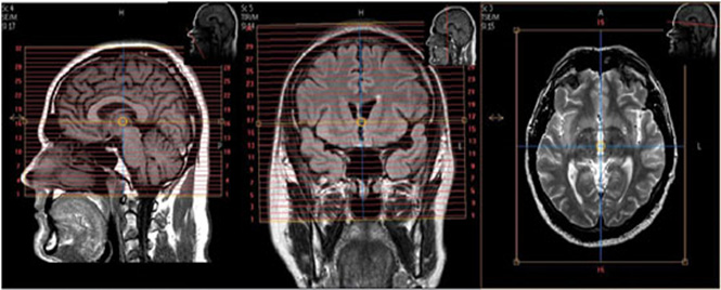
- Landmark at nasion/glabella (±50mm), you must re-landmark in the brain if another body part is scanned first.
- Keep the 3D box as straight as possible.
- No patients with shunts or major artifact-causing items.
- Braces are usually okay, if there is not a great deal of motion, keep head tightly padded.
- Kyphosis prop up hips not head coil.
- Keep patient at Isocenter For patients with small heads and long necks or large heads:
- Keep FOV box positioned higher than normal but not beyond ±50mm from glabella
- May need to reduce/enlarge the FOV for the individual (not beyond 24 - 25.6)
- FOV must include all of scalp, nose and chin.
Required Scan Parameters - NeuroQuant System-specific Parameter Details
Following parameters remain consistent:1. Plane – sagittal
2. Mode – 3D
3. T1 weighted - Always
4. Matrix – 192 x 192
5. NEX / NSA – 1
6. Slice thickness – 1.2 mm
7. Spacing – 1.2 mm
8. Number of slices – 160 - 170
9. FOV 24 – 25.6NOTE: Some NeuroQuant parameters vary depending on scanner manufacturer & field strength
NeuroQuant Failures (And How to Avoid Them)
Patient Motion
- Run NeuroQuant sagittal 3D first
- Pad head securely
- Use all motion reduction techniques except changing scan parameters
Artifacts
- Surgical resections, shunts, metal (some are not compatible)
- Incompatible with NeuroQuant
- Tumors > 15cc
- Contrast media - scan Non Contrast
Low SNR
- Follow recommended scan parameters
Tissue contrast Error
- Put saline bags on either side of patient's head
- Sat bands
- Follow recommended scan parameters
Incorrect landmark
- Must landmark at nasion/glabella
- Can be ± 50mm from Nasion - should be as close as possible in all 3 planes
- Re - landmark, if C-spine was done first as part of a double study
Network Issue
- Echo test failure – call your network admin
- Resend series
- Error report “Series is missing slices”
Unsupported MR Sequence
- Delete incorrect series from queue monitor
- Resend Sag 3D T1 series
-
EFFECTIVE IMMEDIATELY AS OF 10/18/2023:
Effective immediately, we will be pushing the 3DT1 and 3DFLAIR images – please see below for the instructions to follow for this:
One of the important changes with this process is to have the priors processed so that MS Quant will be able to determine new or changes in lesions. We’re looking at ways to have this process done before the patient arrives for their MRI, but until that is in place, we need to do the following:
- Check to see if your patient has a prior MS Brain – we only need to process the most recent prior AND only if it was acquired on the same platform type (i.e., Philips vs Siemens). If this is the case, we need to push the prior 3DT1 and 3D FLAIR to the mode “NQ_MS_PLUS” in PACS. Review the tip sheet (below) for instructions for pushing these sequences from EI PACS. THIS MUST BE DONE BEFORE THE NEW IMAGES ARE PUSHED TO NQ. If there is a MS Quant Processing in place (likely to be rare at this point, it would only be in place for exams acquired starting today) this step can be skipped.
- If the priors have been pushed or the patient doesn’t have a most recent prior on the same platform, when you are done with the MRI, you need to push only the 3DT1 and the 3DFLAIR to the “NQ_MS_NEW” node. The old node is still active on all the scanners during this trial, so please make sure you’re sending to the NEW node. Cases sent to the old node will not process and will tie up NQ.
- Check to see if your patient has a prior MS Brain – we only need to process the most recent prior AND only if it was acquired on the same platform type (i.e., Philips vs Siemens). If this is the case, we need to push the prior 3DT1 and 3D FLAIR to the mode “NQ_MS_PLUS” in PACS. Review the tip sheet (below) for instructions for pushing these sequences from EI PACS. THIS MUST BE DONE BEFORE THE NEW IMAGES ARE PUSHED TO NQ. If there is a MS Quant Processing in place (likely to be rare at this point, it would only be in place for exams acquired starting today) this step can be skipped.
-
Sending Neuroquant Images
Step 1. No Post Processing or Reformatting
Do not reformat or do any post-processing to the NeuroQuant Series. This will cause errors in the DICOM tags that can cause NeuroQuant to reject the Images.Step 2. Send Images Immediately to the Appropriate Node
As soon as you acquire the NQ series, send it to the appropriate node. This allows processing time while the study is completed. NQ processing takes about 15-30 minutes- Multiple Sclerosis: Send to NQ_MS.
- Epilepsy: Send to NQ_EPILEPSY
- Dementia: Send to NQ_DEMENTIA
Step 3. Check to make sure the correct report(s) are generated in PACS. If there are errors, email Joe and Wayne.
- NQ_MS. Generates 1 report: Multistructure Atrophy
- NQ_EPILEPSY. Generates 3 reports: Age-Related Atrophy, Multistructure Atrophy, and Triage Brain Atrophy
- NQ_DEMENTIA. Generates 3 reports: Hippocampal Asymmetry, Multistructure Atrophy, and Triage Brain Atrophy
| Plane | Weighting | Mode | Slice (mm) | Gap (mm) | FAT SAT | FOV | MPR | Notes |
|---|---|---|---|---|---|---|---|---|
| SAG | T1 | 3D TFE | 1.2 (160-170 slices) | 0 | no | 24-25.6 | no | Send to NQ_MS. See specific NeuroQuant 3D T1 scan parameters above. |
| AXIAL | T2 | 2D TSE | 4 | 1 | no | 23 | no | Angle to Corpus. Cover at least 1cm above vertex through skull base. Cover entire nose. |
| AXIAL | DWI | EPI | 3 | 0.3 | YES | 23 | no | Angle to Corpus. Cover Skull Base to Vertex. Send only B1000 & ADC. |
| AXIAL | SWI | 3D GRE | 2 | -1 | no | 23 | no | Angle to Corpus. Cover skull base to vertex. Ok to add slices. |
| Contrast Injection | ||||||||
| SAG | T2 FLAIR | 3D IR-TSE | 1 | 0 | YES | 23 | AXIAL, COR | Angle to interhemispheric fissure. Cover ears and nose. Spacing and gap are variable. |
| SAG | T1 | 3D TSE | 1 | 0 | YES | 23 | AXIAL, COR | Angle to interhemispheric fissure. Cover at least 1cm above vertex through skull base. Cover entire nose. |