MR Meeker Achilles Research Protocol
Scan Notes:
- Scan area: bilateral lower extremity
- Axial, Coronal, and Sag T1
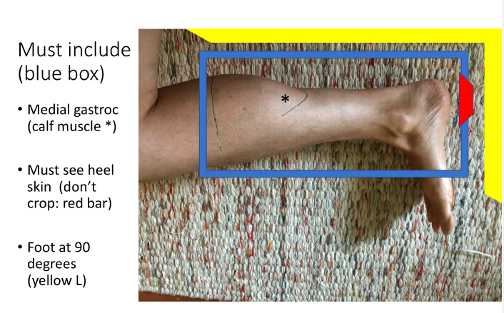
- Refer to images for equipment setup to obtain the best positioning without compressing the heel
- OK to add slices to make sure that you have full coverage
- Do not change FOV or parameters. Run is as built in the scanner
- Cover from upper calf through the heel. Include at least 2cm of air below the heel in the Sag images.
- The heel is more important in the Sag View than the knee, so you can cheat down if needed if the FoV isn't large enough.
- Everything can be angled to the tib/fib.
Last updated: 9/23/2020
Charge as: Bilateral Lower Extremity WO
Scanner preference: MR3 only
Coil: Torso coil
| Sequence | Weighting | Change Parameters? | Change # slices? | Angulation | Notes |
|---|---|---|---|---|---|
| SAG | T1 | NO | yes | Angle to the tib fib. From upper calf through the heel. Bilateral. | Cover from upper calf through the heel. Include at least 2cm of air below the heel in the Sag images. |
| COR | T1 | NO | yes | Angle to the tib fib. From upper calf through the heel. Bilateral. | Cover from upper calf through the heel. |
| AX | T1 | NO | yes | Angle to the tib fib. From upper calf through the heel. Bilateral. | Cover from upper calf through the heel. |