MR Materialise Knee WO Protocol
Ordered by Ortho for surgical planning. When exam completed, email Wayne, Joe, and the leads with the patient name and MRN for upload to the Materialise Surgicase website.
• Do not change the slice thickness, slice increment, matrix, sequence, or FOV.
• Slice number will vary depending on patient size.
Last updated: 8/28/2020
Charge as: Knee WO
Scanner preference: MR2 only
Coil: Knee Coil or torso or flex coil, depending upon patient habitus
-
FREQUENTLY ASKED QUESTIONS
1. MY PATIENT CANNOT STRAIGHTEN THEIR LEG.
Rotation and/or flexion of the knee is allowed up to 20 degrees to make the patient comfortable. Please provide support for the lumbar, knee, and ankle as necessary.2. DOES THE ANKLE NEED TO BE DORSIFLEXED?
No. However, the ankle should be as AP as possible. If necessary, sandbags and other immobilization devices are recommended.3. DO I NEED TO SCAN BILATERAL ANKLES AND HIPS?
Bilateral ankles and/or hips may be scanned, if desired. But, do not increase FOV past the maximum allowed.4. THE CONTRALATERAL KNEE HAS AN IMPLANT/METAL HARDWARE. WHAT DO I DO?
Position the knee as far away from the surgical side as possible; using sandbags to separate the legs and/or cushioning to raise the contra-lateral knee.5. MY PATIENT HAS AN IMPLANT/ METAL HARDWARE IN THE KNEE CAUSING ARTIFACT.
If your patient has an implant or metal hardware in the distal femur or proximal tibia, the artifact may affect critical areas of the scan and the images will be unusable. Please follow-up with the local sales representative or Materialise NV for further assistance; the patient may need to be scanned with CT.6. THE PATIENT HAS A HIP AND/OR ANKLE HARDWARE ON THE SURGICAL SIDE. CAN I STILL SCAN WITH AN MRI?
Yes. Be sure to use a Turbo or Fast Spin Echo with short echo spacing and a high number of echoes. You can also increase the resolution from 256x256 to 512x512, increase the bandwidth, and/or increase the number of averages to 2.7. CAN I USE MORE THAN ONE CONCATENATION ON ANY SCAN?
One package or slab is preferred. It is acceptable for the TR of the ankle or hip to be greater than 800 when necessary to guarantee one slab.8. WHAT PARAMETERS CAN I CHANGE?
• Do not change the slice thickness, slice increment, matrix, sequence, or FOV.
• Slice number will vary depending on patient size.
• TR and TE may vary depending on software.
• Bandwidth can be adjusted as necessary to accommodate for SNR and/or chemical shift.
• Flip Angle can be adjusted to improve SNR.PATIENT PREPARATION
- Discuss the procedure with the patient to ensure they understand the table will move during scanning.
- Instruct patient not to move during any part of the scanning sequence. Patient movement will alter the relative alignment of the joints and invalidate the scan.PATIENT POSITIONING
- Knee coil preferred, but if your patient does not fit, ok use the torso or flex coil.
- Position the patient's knee as close to ISO-center as possible.
- Position Apex of the patella at the center of the coil
- Position the patient so the side of interest is as close to the center of the table, left to right, as possible.
- Rotation of the knee is allowed up to 20 degrees to make the patient comfortable as long as the knee remains in ISO-center and the knee coil is properly placed. Do not hyper-flex the knee.
- Ensure the ankle is scanned on the same plane or slightly lower than the knee joint: the protocol allows for up to a 20 degree flexion of the knee joint.
- Immobilize the leg using sandbags and straps to restrict external rotation of the knee and stabilize the leg.
- Lumbar support is recommended to relieve back pain while the legs are extended.
- Landmark at the center of the coilIMAGING NOTES
- The offsets defined in the following procedure are approximations only. Enter the table coordinates for each joint as precisely as possible; the actual offset will be based on the patient’s anatomy, not a default value.
- Multiple localizers are acceptable, as long as the patient is not re-positioned or re-landmarked.
HIGH-RES SAG KNEE
Knee Coil or torso or flex coil, depending upon patient habitus
| SEQUENCE | Ok to Angle? | Coverage | Slice Thickness | Matrix | Sequence | TE | Bandwidth | FOV | TR | Flip Angle | Fat Saturation | Notes |
|---|---|---|---|---|---|---|---|---|---|---|---|---|
| Localizer | NO ANGLE | |||||||||||
| HIGH RES SAG | DO NOT ANGLE | Include 10 cm of femur and 10 cm of tibia to include femoral condyles and tibial plateau and tibia tuberosity. Must have good signal. | 2mm with Overcontiguous Slices ON | ACQ matrix 256 x 256 with Reconstruction Matrix 512 | 3D WATSc | Set TE to Shortest "In Phase" | non-select | 200-250 mm | 10-20 | 10-25 | Pro Set | Technique T1-FFE. Clear/Sense is allowed. Do not use No Phase Wrap, Oversampling, or Fold-Over Suppression |
LOW RES ANKLE (BODY COIL)
Remove the knee coil and landmark at the malleoli of the ankle. Patient must not move when removing knee coil. Bilateral ankles not required. The low-resolution series is used to calculate the length of the femur and tibia, as well as the full alignment of the entire limb. This is done by tracking the table position/table coordinates of each separate joint scan.
| SEQUENCE | Ok to Angle? | Coverage | Slice Thickness | Matrix | Sequence | TE | Bandwidth | FOV | TR | Flip Angle | Fat Saturation | Notes |
|---|---|---|---|---|---|---|---|---|---|---|---|---|
| Localizer | NO ANGLE | |||||||||||
| LOW RES AXIAL ANKLE (NON-FAT SAT) | DO NOT ANGLE | Cover from above the malleoli to the mid-calcaneus. | 5mm | 256 x 256 | 2D TSE | 20 | non-select | 260mm | - | - | NONE | NSA = 1 |
LOW RES KNEE (BODY COIL)
Move table to scan the knee using superior offset ~300-400mm (this amount will vary per patient). Do not re-landmark.
| SEQUENCE | Ok to Angle? | Coverage | Slice Thickness | Matrix | Sequence | TE | Bandwidth | FOV | TR | Flip Angle | Fat Saturation | Notes |
|---|---|---|---|---|---|---|---|---|---|---|---|---|
| Localizer | NO ANGLE | |||||||||||
| LOW RES SAG KNEE (FAT SAT) | DO NOT ANGLE | Include 10 cm of femur and 10 cm of tibia. | 5mm | Set reconstruction matrix to 256 | 3D WATSc | Set TE to Shortest ?In Phase? | non-select | 260mm | 10-20 | 10-25 | Pro Set | Technique T1-FFE. Clear/Sense is allowed. Do not use No Phase Wrap, Oversampling, or Fold Over Suppression |
LOW RES HIP (BODY COIL)
Move table to scan the knee using superior offset ~300-400mm (this amount will vary per patient). Do not re-landmark. Bilateral hips not required.
| SEQUENCE | Ok to Angle? | Coverage | Slice Thickness | Matrix | Sequence | TE | Bandwidth | FOV | TR | Flip Angle | Fat Saturation | Notes |
|---|---|---|---|---|---|---|---|---|---|---|---|---|
| Localizer | NO ANGLE | |||||||||||
| LOW RES AXIAL HIP (NON-FAT SAT) | DO NOT ANGLE | Cover from anterior superior iliac spine to pubic symphysis. Cover the femoral head and neck. | 5mm | 256 x 256 | 2D TSE | 20 | non-select | 360mm | - | - | NONE |
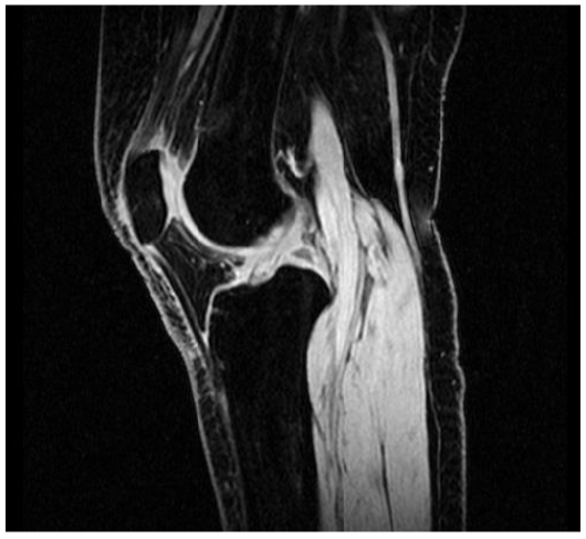
HIGH-RES SAGITTAL
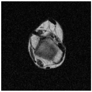
LOW-RES AXIAL ANKLE
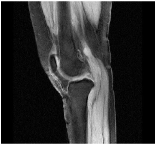
LOW-RES SAGITTAL KNEE
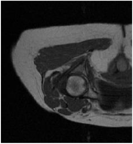
LOW-RES AXIAL HIP