MR Liver Mass W/WO BODY Protocol
Scan Notes
Last updated: 1/31/24
Charge as: Abdomen W/WO
Scanner preference: 1.5T or 3T
Coil: Torso Coil
- Void before exam
- Send ADC maps
- FOV: do not include patient’s arms
Breath Holds:
- Scan on expiration.
- Monitor that patient is breath-holding. Breathe the patient slowly so they have time to follow instructions. Do not start scan until the patient has stopped breathing.
- Give 2L O2 if it will help with breath-holds UNLESS PATIENT HAS COPD OR ANOTHER REASON NOT TO GIVE O2.
| Plane | Weighting | Mode | Slice | Gap | FAT SAT | FOV | Notes |
|---|---|---|---|---|---|---|---|
| Cor | T2 | SSFSE BH | 7mm | 1mm | none | Liver through bottom of kidneys | Ensure liver is well-centered in coil to ensure good signal at dome. |
| Axial | T1 | Dual Echo SPGR BH | 5mm | 0.5mm | none | Entire liver | May be separated into overlapping stacks if patient cannot breath-hold. Do not interleave images. Okay to use 6mm slice thickness with 1mm gap on MR1 and CHMR2. |
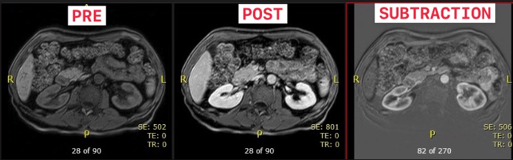
| Axial | T1 | 3D mDixon pre BH | - | - | Yes | Entire liver | Ensure quality before contrast injection |
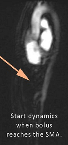
| Power Inject Contrast | Bolus Track | Trigger when bolus reaches SMA. | |||||
| Axial | T1 | 3D mDixon x3 phases BH | - | - | Yes | Entire liver | Exact parameters as the pre-mDixon. |
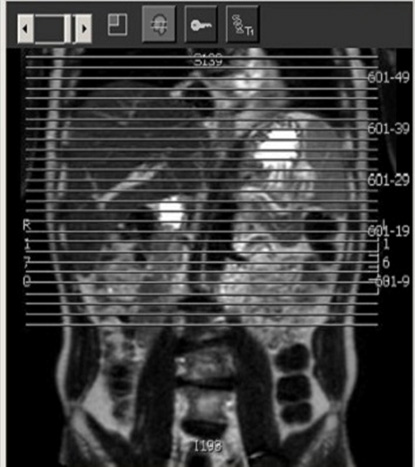
| COR | T1 | 3D mDixon 3 min post | - | - | Yes | Diaphragm to Aortic bifurcation | Perform @ 3 minutes post contrast. Abdominal wall to abdominal wall |
| Axial | T1 | 3D mDixon BH | - | - | Yes | Entire liver | Timed exactly 4 mins post-injection. |
| Axial | T2 | TSE RT | 5mm | 0.5mm | SPAIR | Entire liver | Okay to use 6mm slice thickness with 1mm gap on MR1 and CHMR2. |
| Axial | T2 | DWI | 7mm | 1mm | Liver through bottom of kidneys | ||
| Axial | T2 | SSFSE RT | 5mm | 0.5mm | Yes | Entire liver | Optional sequence if T2 SPAIR is poor quality. Okay to use 6mm slice thickness with 1mm gap on MR1 and CHMR2. |