MR Female Pelvis / Soft Tissue Pelvis W/WO BODY Protocol
Scan Notes
Last updated: 4/12/19
Charge as: Pelvis W/WO
Scanner preference: 1.5T
Coil: Torso Coil
- Void before exam
- Send ADC maps
| Plane | Weighting | Mode | Slice | Gap | FAT SAT | FOV | Notes |
|---|---|---|---|---|---|---|---|
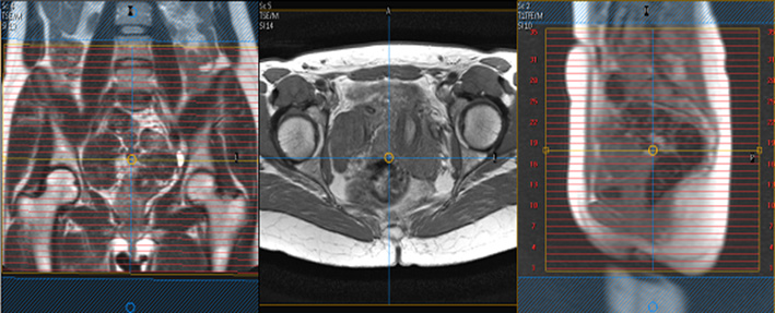
| COR | T2 | SSTSE BH | 5mm | 1mm | N | top of kidneys to pelvis | Scan sacrum to anterior abdominal wall. CONFIRM GOOD COIL PLACEMENT. Large FOV to include kidneys. Pelvic pathology is often related to renal pathology |
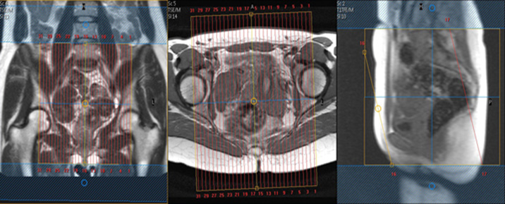
| AXIAL | T1 | TSE | 5mm | 1mm | N | 20-24mm, fit to patient | Scan iliac crest to perineum. Freq A-P to avoid bowel motion ghosting into uterus and bladder. If there is a pelvic mass, please scan to include the whole mass. |
| AXIAL | T2 | TSE | 5mm | 1mm | N | 20-24mm, fit to patient | Scan iliac crest to perineum. Same parameters as AX T1 TSE. Freq A-P to avoid bowel motion ghosting into uterus and bladder. |
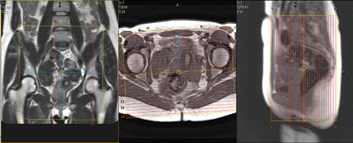
| SAG | T2 | TSE | 5mm | 1mm | N | 24mm, fit to patient | Scan mid-femoral head to mid-femoral head. Freq A-P. Consider using an anterior Sat band if lots of abdominal wall motion. |
| COR | T2 | TSE FS | 5mm | 1mm | Y | 20-24mm, fit to patient | Scan sacrum to anterior abdominal wall. Freq A-P. |
| Optional AX Oblique | T2 | TSE | 5mm | 1mm | N | 20-24mm, fit to patient | Optional Sequence for Uterine/Mullerian anomaly.Scan uterus to perineum. Slices should be along the length of the uterus.CALL Rad FOR PLANNING! |
| Optional COR Oblique | T2 | TSE | 5mm | 1mm | N | 20-24mm, fit to patient | Optional Sequence for Uterine/Mullerian anomaly. Include Uterus, Cervix and vagina. Slices should be along the short axis of the uterus. CALL Rad FOR PLANNING! |
| AXIAL PRE | T1 | 3D thrive high resolution pre-contrast | Y | Match AX TSE T2 Pelvis | HIGH RESOLUTION THRIVE.Not a dynamic sequence. | ||
| Hand Inject Contrast | |||||||
| AXIAL POST | T1 | 3D thrive high res | Y | Match high resolution precontrast THRIVE | HIGH RESOLUTION THRIVE Not a dynamic sequence. | ||
| SAG | T1 | 3D thrive high res | Y | Match SAG TSE T2 Pelvis | HIGH RESOLUTION THRIVEs. | ||
| COR | T1 | 3D thrive high res | Y | Match COR TSE FS T2 Pelvis | HIGH RESOLUTION THRIVEs. | ||
| AXIAL | T2 | DWI | 5mm | 1mm | SPIR | Match AX TSE T2 Pelvis | Trigger & track. Free-breathing sequence, so please position slices accordingly. B=0, 500, 1000. |