MR Elbow Arthrogram WO MSK Protocol
Scan Notes:
Patient will come down from X-Ray after the injection. Images do not need to be checked by the radiologist unless there are questions or concerns.
Last updated: 4/8/19
Charge as: Elbow Arthrogram WO
Scanner preference: 3T
Coil: Knee or Flex Coil
| Plane | Weighting | Mode | Slice | Gap | FAT SAT | FOV | Notes |
|---|---|---|---|---|---|---|---|
| AXIAL | T2 SPAIR | TSE | 3mm | 0.5mm | SPAIR | 14cm | 4cm Above/ Below Elbow Joint |
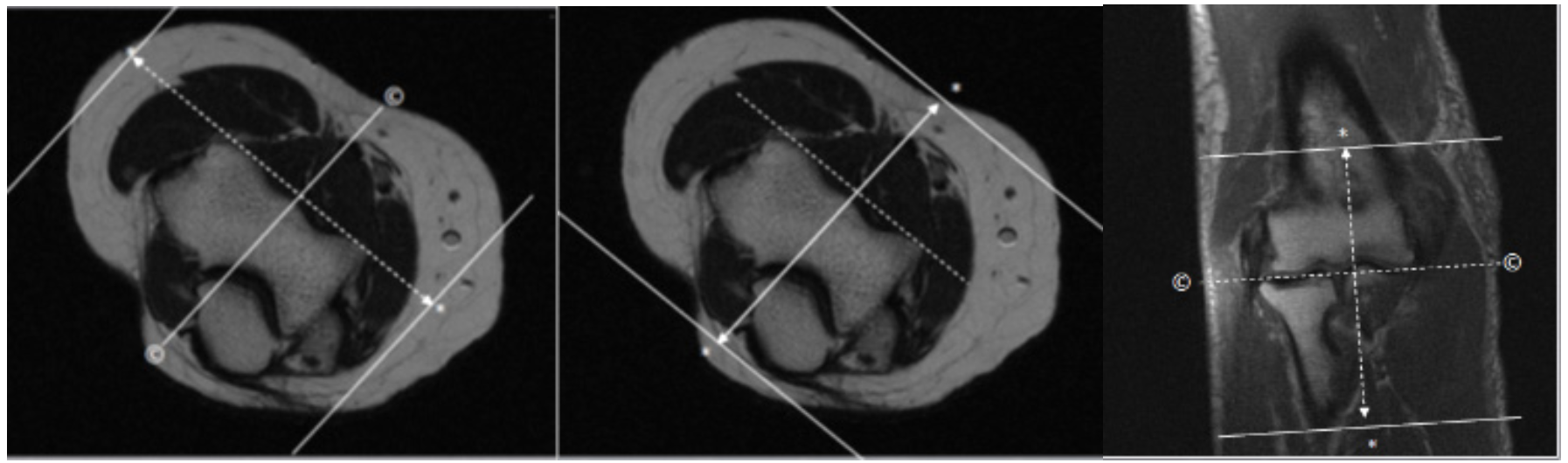
| SAG | T2 STIR | TSE | 3mm | 0.5mm | STIR | 14cm | Angle to Distal Humeral Condyles |
| COR | T2 STIR | TSE | 3mm | 0.5mm | STIR | 14cm | Angle to Distal Humeral Condyles |
| COR | T1 | TSE | 3mm | 0.5mm | None | 14cm | Angle to Distal Humeral Condyles |
| AX | T1 SPIR | TSE | 3mm | 0.5mm | SPIR | 14cm | Angle to Distal Humeral Condyles |
| SAG | T1 SPIR | TSE | 3mm | 0.5mm | SPIR | 14cm | Angle to Distal Humeral Condyles |
| COR | T1 SPIR | TSE | 3mm | 0.5mm | SPIR | 14cm | Angle to Distal Humeral Condyles |