MR Liver Mass with MRCP WO BODY Protocol
Scan Notes:
Last updated: 1/31/2024
Charge as: Abdomen and MRCP WO
Scanner preference: 1.5T or 3T
Coil: Torso Coil
- Void before exam
- FOV: do not include patient’s arms
Breath Holds:
- Scan on expiration unless patient is having difficulty breathing.
- Monitor that patient is breath-holding. Breathe the patient slowly so they have time to follow instructions. Do not start scan until the patient has stopped breathing.
- Give 2L O2 if it will help with breath-holds UNLESS PATIENT HAS COPD OR ANOTHER REASON NOT TO GIVE O2.
| Plane | Weighting | Mode | Slice | Gap | FAT SAT | FOV | Notes |
|---|---|---|---|---|---|---|---|
| Cor | T2 | SSFSE BH | 7mm | 1mm | No | Liver through bottom of kidneys | Ensure liver is well-centered in coil to ensure good signal at dome. |
| Axial | T1 | Dual Echo SPGR BH | 5mm | 0.5mm | No | Entire liver | May be separated into overlapping stacks if patient cannot breath-hold. Do not interleave images. Okay to use 6mm slice thickness with 1mm gap on MR1 and CHMR2. |
| Axial | T2 | bTFE BH | 4mm | -2mm | No | Perform 3 overlapping stacks. Do not interleave. | |
| Axial | T1 | 3D mDixon BH | - | - | Yes | Entire liver | |
| Axial | T2 | TSE RT | 5mm | 0.5mm | SPAIR | Entire liver | Okay to use 6mm slice thickness with 1mm gap on MR1 and CHMR2. |
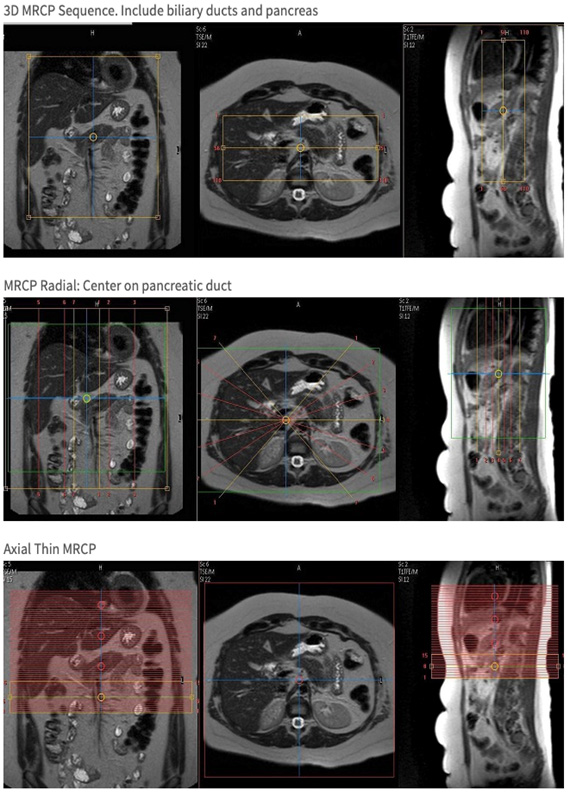
| Cor | T2 | 3D MRCP | 1.4mm | Yes | Bottom 2/3 of liver through bottom of pancreas | Please use navigator trigger & track. | |
| Cor | T2 | Radial MRCP | 40mm | Yes | Liver through bottom of kidneys | Please use navigator trigger & track. | |
| Axial | T2 | DWI | 7mm | 1mm | SPIR | Liver through bottom of kidneys | Trigger & Track |
| COR | T1 | 3D mDixon | - | - | Yes | Cover diaphragm to aortic bifurcation, abdominal wall to abdominal wall | |
| Axial | T2 | SSFSE RT | 5mm | 0.5mm | Yes | Entire liver | Optional sequence if T2 SPAIR is poor quality. Okay to use 6mm slice thickness with 1mm gap on MR1 and CHMR2. |