MR Adult Female Pelvis for Cervical Cancer Staging W/WO BODY Protocol
Last updated: 12/8/2023
Charge as: Pelvis W/WO
Scanner preference: 1.5T or 3T
Coil: Torso Coil
- This is the Cervical Cancer protocol, except obliques are relative to the endocervical canal rather than the endometrial canal.
- Send ADC maps and subtractions
- FOV: do not include patient’s arms
| Plane | Weighting | Mode | Slice | Gap | FAT SAT | FOV | Notes |
|---|---|---|---|---|---|---|---|
| COR | T2 | SSTSE BH | 5mm | 1mm | N | Top of kidneys → pelvis. Sacrum → anterior abdominal wall | CONFIRM GOOD COIL PLACEMENT. Large FOV to include kidneys. Pelvic pathology is often related to renal pathology. |
| SAG | T2 | TSE | 4mm | 1mm | N | 200-240 mm. Acetabulum → Acetabulum | Consider using an anterior Sat band if lots of abdominal wall motion. If there is a pelvic mass, please scan to include the whole mass. Matrix 256 x 256 |
| AXIAL OBL | T2 | TSE | 3mm | 0.5mm | N | 200-240 mm/ Fit to Patient. Uterus → rectum | Perpendicular to the endocervical canal. Use all planes to obtain true axial of the endocervical canal (see images below). Resulting image should be a true “donut.” Matrix 512 x 256-512. Freq A-P. |
| AXIAL OBL | T2 | DWI | 4mm | 1mm | SPIR | Match AX OBLIQUE | Trigger & track. Free-breathing sequence, so please position slices accordingly. B=0, 500, 1000. |
| AX | T1 | TSE | 5mm | 1mm | N | 20-24 mm/Fit to Patient. Top L5 → perineum | Freq A-P to avoid bowel motion ghosting into uterus and bladder. If there is a pelvic mass, please scan to include the whole mass. |
| SAG | T1 | 3D THRIVE precontrast | -- | -- | Y | 20-24 mm. Acetabulum → Acetabulum | Non-high resolution THRIVEs. |
| Dynamic Contrast Injection | |||||||
| SAG dynamic (40 seconds, 1 minute, 90 seconds) | T1 post | 3D THRIVE post contrast BH | -- | -- | Y | 20-24 mm. Acetabulum → Acetabulum | NON-high resolution THRIVEs. Perform at 40s, 60s, 90s post contrast. |
| 3 minutes post AX | T1 | 3D THRIVE post contrast BH | -- | -- | Y | 20-24 mm/Fit to Patient. Top L5 → perineum | NON-high resolution THRIVEs. Perform at 180s post contrast |
| AX | T2 post | TSE | 4mm | Y | Top L5 → perineum |
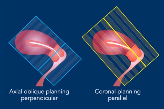
Sequence Planning
- The MR sequences are planned relative to the long axis of the cervical canal.
- The axial plane is perpendicular to the long axis of the cervical canal.
- The coronal plan is parallel to the long axis of the cervical canal.
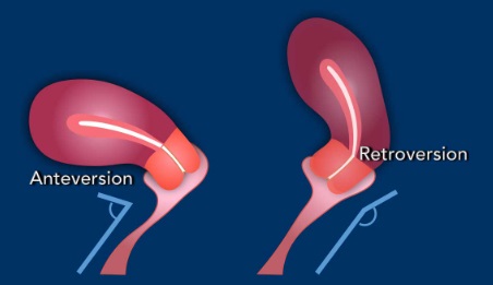
Pitfall: variations in cervical anatomy
The position of the cervical canal needs to be taken into account and the perpendicular and parallel MRI sequences need to be planned accordingly.
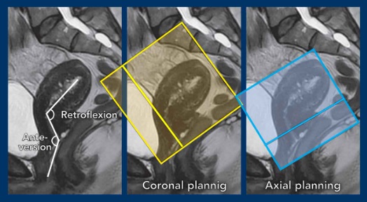
Example showing how flexion, and in particular version impact sequence planning.
- In this case there is anteversion of the cervix and retroflexion of the uterus.
- Remember that in cervical cancer, the axial sequences are planned perpendicular to the cervical canal.
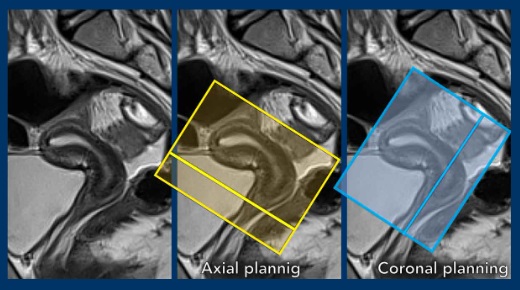
Another example showing the cervix in retroversion and the uterus in anteflexion.
See how this variation in position impacts the corresponding sequence planning.