MRI Toe WO or W/WO MSK Protocol
Scan Notes:
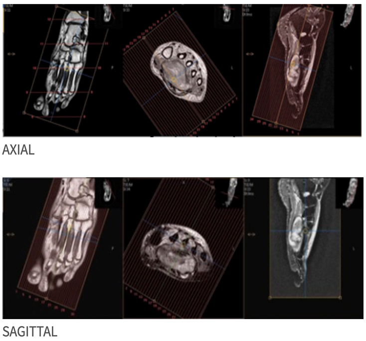
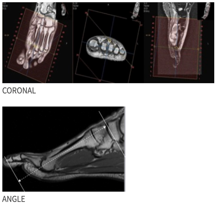
- Angle to Metatarsals
- Cover 3cm proximal to MTP joint thru tip of toe
- Cover area of interest and 1-2 toes on either side of affected toe
- If Sag T2 Fat Sat has poor fat suppression, consider STIR (TE = 40-45) instead
Last updated: 4/8/19
Charge as: Toe WO or W/WO
Scanner preference: 1.5T
Coil: Torso Coil
| Plane | Weighting | Mode | Slice | Gap | FAT SAT | FOV | Notes |
|---|---|---|---|---|---|---|---|
| AXIAL | T2 SPAIR | TSE | 3mm | 0.5mm | SPAIR | see below | Cover 3cm proximal to MTP joint thru tip of toe |
| SAG | T2 SPAIR | TSE | 3mm | 0.5mm | SPAIR | Cover area of interest and 1-2 toes on either side of affected toe | Cover 3cm proximal to MTP joint thru tip of toe. If poor fat suppression, consider STIR (TE = 40-45) instead |
| SAG | T1 | TSE | 3mm | 0.5mm | None | Cover area of interest and 1-2 toes on either side of affected toe | Cover 3cm proximal to MTP joint thru tip of toe |
| COR | T1 | TSE | 3mm | 0.5mm | None | see below | Cover 3cm proximal to MTP joint thru tip of toe |
| COR | T2 SPAIR | TSE | 3mm | 0.5mm | SPAIR | see below | Cover 3cm proximal to MTP joint thru tip of toe |
| AX PRE(if giving contrast for infection/osteo) | T1 | TSE | 3mm | 0.5mm | NONE | see below | Same as AX T2 FS |
If ordered with contrast
| Plane | Weighting | Mode | Slice | Gap | FAT SAT | FOV | Notes |
|---|---|---|---|---|---|---|---|
| AXIAL | T1 SPIR | TSE | 3mm | 0.5mm | SPIR | Same as pre | Same as pre |
| SAG | T1 SPIR | TSE | 3mm | 0.5mm | SPIR | Same as pre | Same as pre |
| COR | T1 SPIR | TSE | 3mm | 0.5mm | SPIR | Same as pre | Same as pre |