MRI Sacroiliitis/SI Joints/Sacrum WO or W/WO MSK Protocol
Scan Notes:
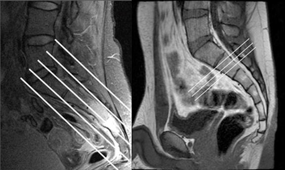
Angle OBLIQUE to sacrum
If hardware is present:
- Do Axial and Coronal STIR instead of fat-sat mid-TE
- If with Contrast, do non-fat-sat T1 post-contrast
Last updated: 4/8/19
Charge as: Pelvis WO or W/WO
Scanner preference: 1.5T or 3T
Coil: Torso Coil
| Plane | Weighting | Mode | Slice | Gap | FAT SAT | FOV | Notes |
|---|---|---|---|---|---|---|---|
| AXIAL OBL | T1 (TE=min) | TSE | 4-5 mm | 1 mm | None | Cover bilateral SI Joints, sacrum and coccyx. | Angle perpendicular to Oblique Coronals (perpendicular to the sacrum). |
| AXIAL OBL | T2 SPAIR | TSE | 4-5 mm | 1 mm | SPAIR | Cover bilateral SI Joints, sacrum and coccyx. | Angle perpendicular to Oblique Coronals (perpendicular to the sacrum). |
| SAG | T2 SPAIR | TSE | 3-4 mm | 0.5 mm | SPAIR | Cover entire sacrum and SI joints from L4-5 to tip of the coccyx. No need for entire pelvis. | Include lateral femoral head margins |
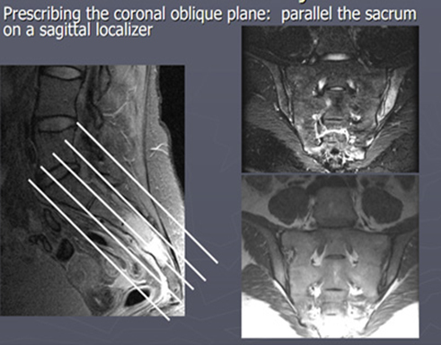
| COR OBL | T1 (TE=min) | TSE | 3-4 mm | 0.5 mm | None | Cover L4-5 to tip of the coccyx through SI Joints and Sacrum. No need for entire pelvis. | Include lateral femoral head margins. Angle parallel to long axis of sacrum (perpendicular to Axial Obl). |
| COR OBL | STIR (TE=70msec) | TSE | 3-4 mm | 0.5 mm | STIR | Cover L4-5 to tip of the coccyx through SI Joints and Sacrum. No need for entire pelvis. | Include lateral femoral head margins. Angle parallel to long axis of sacrum (perpendicular to Axial Obl). |
| SAG PRE (if giving contrast for infection/osteo) | T1 | TSE | 3-4 mm | 0.5 mm | NONE | Same as SAG T2 FS | Same as SAG T2 FS |
If Ordered with Contrast
| Plane | Weighting | Mode | Slice | Gap | FAT SAT | FOV | Notes |
|---|---|---|---|---|---|---|---|
| COR OBL | T1 THRIVE | TSE | 3-4 mm | 0.5 mm | THRIVE | Same as pre | Same as pre |
| AXIAL OBL | T1 SPIR | TSE | 4-5 mm | 0.5 mm | SPIR | Same as pre | Same as pre |