MR Stroke Brain WO Neuro Protocol
Last updated: 06/30/2023
Charge as: Brain WO
Scanner preference: 1.5T or 3T
Coil: Head
| Plane | Weighting | Mode | Slice (mm) | Gap (mm) | FAT SAT | FOV (cm) | MPR | Notes |
|---|---|---|---|---|---|---|---|---|
| AXIAL | DWI | EPI | 3 | 0.3 | YES | 23 | no | Angle to Corpus. Cover Skull Base to Vertex. Send only B1000 & ADC. |
| COR | DWI | EPI | 3 | 0.3 | YES | 23 | no | Send only B1000 & ADC. |
| AXIAL | T2* | GRE | 4 | 1 | YES | 23 | no | Angle to Corpus. |
| SAG | T1 | TSE | 4 | 1 | no | 24-25.6 | no | Scalp to scalp. |
| AXIAL | T2 | TSE | 4 | 1 | no | 23 | no | Angle to Corpus. |
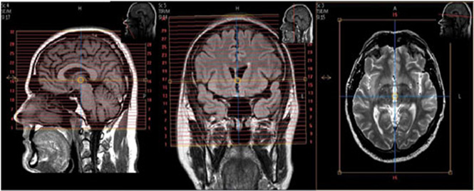
| SAG | T2 FLAIR | 3D IR-TSE | 1 | 0 | YES | 23 | AXIAL, COR OBLIQ | Angle to interhemispheric fissure. Cover ears and nose. Spacing and gap are variable. |
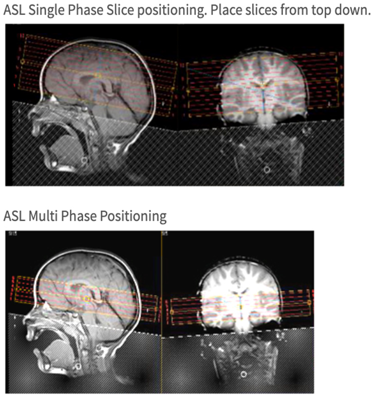
| AXIAL | ASL Single Phase | FFE EPI | 6 | 1 | no | 23 | no | Place from top down. Don't send raw data to PACS. |
| AXIAL | ASL Multi Phase | FFE EPI | 6 | 1 | no | 23 | no | See screen capture for slice placement. Archive raw data to DVD. |