MR Spectroscopy - Single Neuro Protocol
Scan Notes:
The radiologist will plan the spect voxels.
Typically they will want one on whatever lesion they are looking at, and one in normal healthy white matter.
Voxel sizes: maximum 20 x 20 x 20, but you can go as small as 10 x 10 x 10.
Make every effort possible to avoid placing any part of the voxelss inside CSF, bone, or air.
You may turn the voxel boxes in any direction to help achieve this.
It is useful to run an additional COR T2 to help plan the voxels.
Last updated:3/21/2019
Charge as: Usually added on to a Neuro Brain exam. No additional charge needed.
Scanner preference: 3T only
Coil: Head
Post processing
- Once the spect scan has completed
- Right mouse click on the spect and choose "Spectroview" sequence
- Click the icon link on the far left on the upper part of the screen.
- Click on "LongTeBrain" and then click "OK"
- Choose "Spectrum Phase Adjustment" so the checkmark is visible in the box. Do not click any other options.
- Click "RUN"
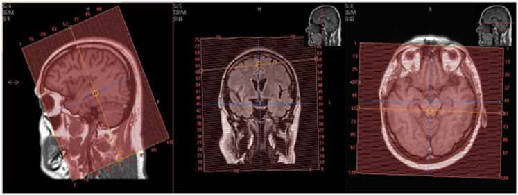
- The next screen will show you three planes. Scroll through the images in each box to MAKE SURE YOU SEE THE RED BOX IN EVERY VIEW
- Scroll through the images of the brain with your mouse until you can see the RED VOXEL BOX in every view.
- Make a screen capture of this screen and send to PACS.