MR Framed Brain WO Pre and Post DBS Neuro Protocol
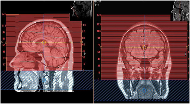
Scan Notes: The patient will arrive with the MD and will have the stereotactic grid in place. The frame is located in MRI scanner room 1. This attaches to the patient’s framed head and seats inside the T/R coil. No angles, no parameter changes! The MD with the patient will tell you which scans to run.
Last updated:3/21/2019
Charge as: Brain WO
Scanner preference: 1.5T only
Coil: T/R Head coil only
| Plane | Weighting | Mode | Slice | Gap | FAT SAT | FOV | Notes |
|---|---|---|---|---|---|---|---|
| AXIAL | 3D T1 FFE | 1cm below Skull Base to 1cm above Vertex. You must add enough slices to capture the fiducials that are inside the frame. | |||||
| AXIAL | Flair Stereo | 1cm below Skull Base to 1cm above Vertex. You must add enough slices to capture the fiducials that are inside the frame. | |||||
| AXIAL | STIR/TSE | 1cm below Skull Base to 1cm above Vertex. You must add enough slices to capture the fiducials that are inside the frame. | |||||
| AXIAL | T2/ TSE | 1cm below Skull Base to 1cm above Vertex. You must add enough slices to capture the fiducials that are inside the frame. | |||||
| AXIAL | T2 Vista | 1cm below Skull Base to 1cm above Vertex. You must add enough slices to capture the fiducials that are inside the frame. |