MR Cine WO Protocol
Scan Notes: Make sure you have excellent signal at the base of the skull for CSP cine studies.
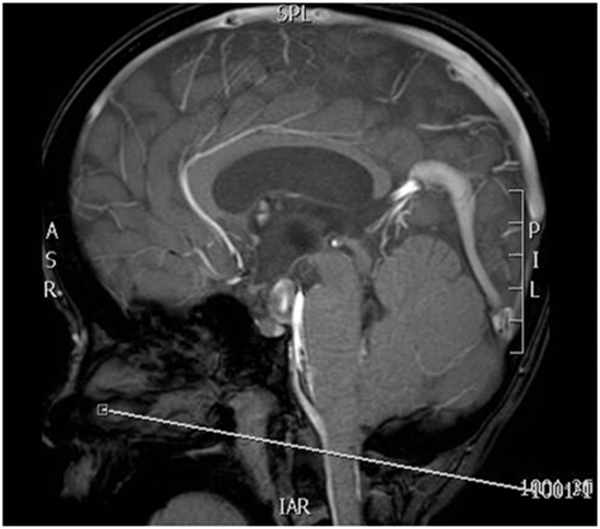
Page the radiologist to plan and place the axial cine sequence. The positioning will depend on the patient anatomy, but it will generally be in this area (see image below). Feel free to set it up and send the rads a screenshot of what you have done. Images must be checked.
Last updated:2/14/13
Charge as: “With Cine” for either CSP, Brain or LSP studies
Scanner preference: 1.5T or 3T
Coil: Head cine = Head coil; CSP cine = NV or Spine coil; LSP = Spine coil
| Plane | Weighting | Mode | Slice | Gap | FAT SAT | FOV | Notes |
|---|---|---|---|---|---|---|---|
| COR | T2 TSE | TSE | 4mm | 1mm | None | 20cm | Whole Brain |
| AXIAL | T2 TSE | TSE | 4mm | 1mm | None | 20cm | Whole Brain |
| SAG | T2 TSE | TSE | 4mm | 1mm | None | 20cm | Whole Brain |
| SAG CINE | PCA- VENC 5 | FFE | 10mm | 0mm | None | 18cm | MSP- Centered on Foramen Magnum |
| SAG CINE | PCA- VENC 12 | FFE | 10mm | 0mm | None | 18cm | MSP- Centered on Foramen Magnum |
| AXIAL CINE | PCA- VENC 5 | FFE | 10mm | 0mm | None | 18cm | To be placed with guidance by the RAD |
| AXIAL CINE | PCA- VENC 12 | FFE | 10mm | 0mm | None | 18cm | To be placed with guidance by the RAD |
Contrast injection
| Plane | Weighting | Mode | Slice | Gap | FAT SAT | FOV | Notes |
|---|---|---|---|---|---|---|---|
| Coronal | FLAIR | TSE | 4 mm | 1 mm | None | 23 cm | Frontal through Occipital Bone |
| Axial | T1 | TSE | 4 mm | 1 mm | None | 23 cm | Angle to Corpus- Skull Base to Vertex |
| Coronal | T1 FAT SAT | TSE | 4 mm | 1 mm | SPIR | 23 cm | Frontal through Occipital Bone |