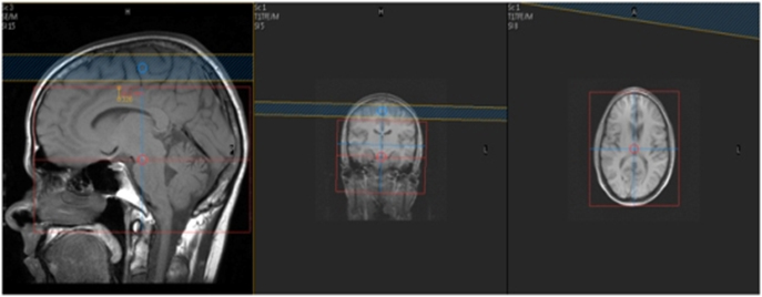
MR Brain Vasculitis WWO + MRA WO Neuro Protocol
Last updated:2/14/13
Charge as: Brain WWO and MRA WO
Scanner preference: MRI1, MRI2, MRI3 or MRI4
Coil: Head
| Plane | Weighting | Mode | Slice | Gap | FAT SAT | FOV | Notes |
|---|---|---|---|---|---|---|---|
| SAG | T1 | TSE | 4mm | 1mm | None | S | Scalp to Scalp |
| SAG | T1 | FLAIR | 4mm | 1mm | None | 23cm | Scalp to Scalp. |
| AXIAL | T2 | TSE | 4mm | 1mm | None | 23cm | Angle to Corpus- Skull Base to Vertex |
| AXIAL | T1 | TSE | 4mm | 1mm | None | 23cm | Angle to Corpus- Skull Base to Vertex |
| AXIAL | T2* GRE | GRE | 4mm | 1mm | None | 23cm | Angle to Corpus- Skull Base to Vertex |
| AXIAL | DWI 2mm Voxel | SE EPI | 3mm | 0.3mm | SPIR | 23cm | Angle to Corpus- Skull Base to Vertex |
| AXIAL | FLAIR | TSE | 4mm | 1mm | None | 23cm | Angle to Corpus- Skull Base to Vertex |
| COW | 3D TOF | 3D FFE | 1mm | 0mm | None | 20cm | MIP COW, Right, Left and Posterior |
Contrast injection
| Plane | Weighting | Mode | Slice | Gap | FAT SAT | FOV | Notes |
|---|---|---|---|---|---|---|---|
| AX DSC | 3D T1 FFE | EPI | 60 | None | 20cm | ||
| Notes: | Scan for 8-10 Seconds then begin contrast injection @ 5ml/s15ml Saline chaserASL Single Phase Slice Positioning. Place slices from top down. | ||||||
| COR | FLAIR | TSE | 4mm | 1mm | None | 23cm | Frontal through Occipital Bone |
| SAGITTAL | T1 flair(T1 NOT T2 weighted). High Resolution | TSE | 3mm | 0mm | SPIR (mild) | 16-18cm | (ok to crop a few cm on either side); center over COW MCAs. Be sure coverage includes the proximal COW to the MCA bifurcation on each side. Can reduce slices to around 24 to keep scan time under 7 minutes.Turn flow compensation on |
| AXIAL | T1 flair(T1 NOT T2 weighted). High Resolution | TSE | 3mm | 0mm | SPIR (mild) | 16-18cm | Angle to Corpus center over middle cerebral artery (MCA). Be sure coverage includes the proximal COW to the MCA bifurcation on each side. Can reduce slices to around 24 to keep scan time under 7 minutes. Turn flow compensation on |
| AXIAL | T1 | TSE | 4mm | 1mm | None | 23cm | Angle to Corpus- Skull Base to Vertex |
| COR | T1 FAT SAT | TSE | 4mm | 1mm | SPIR | 23cm | Frontal through Occipital Bone |
| SAG | T1 | TSE | 4mm | 1mm | None | 23cm | Scalp to Scalp. Match to SAG T1 FLAIR PRE |
