MR Ankle WO MSK Protocol
Scan notes:
For Ligament Tear, Trauma, AVN, Stress F/x, Plantar Fasciitis:
If sag T2 Fat Sat has poor fat suppression, consider STIR (TE=40) instead.
If hardware is present:
- Do AX non-fat-sat mid-TE instead of fat-sat
- Keep SAG STIR
- Do COR STIR instead of fat-sat mid-TE
- If ordered with Contrast, do non-fat-sat T1 post-contrast
Last updated: 4/8/19
Charge as: Ankle WO
Scanner preference: 3T only
Coil: Ankle Coil
| Plane | Weighting | Mode | Slice | Gap | FAT SAT | FOV | Notes |
|---|---|---|---|---|---|---|---|
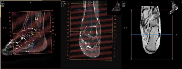
| AXIAL | T2 SPAIR | TSE | 4mm | 1mm | SPAIR | 15cm | Bottom of heel to above malleolus |
| SAG | T2 STIR | TSE | 3mm | 1mm | STIR | 15cm | Angle to Calcaneus |
| SAG | T1 | TSE | 3mm | 1mm | None | 15cm | Angle to Calcaneus |
| COR | T1 | TSE | 3mm | 1mm | None | 15cm | Include Tarsals |
| COR | T2 SPAIR | TSE | 3mm | 1mm | SPAIR | 15cm | Include Tarsals |
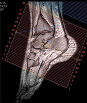
| OptionalAxial ObliquePlantar Flexion | T2 SPAIR | TSE | 4mm | 1mm | SPAIR | 15cm | Performed if requested.Angle to short axis of tarsals. Cover entire tarsals. |
| OptionalAxial ObliquePlantar Flexion | PD | TSE | 4mm | 1mm | None | 15cm | Perform if requested.Angle to short axis of tarsals. Cover entire tarsals. |