MR Thumb WO and W/WO Protocol
Revised - 2/22/19
- 3T scanner only
- Use flex or head coil
- Use rest slab proximal to wrist to reduce flow and pulsatile motion
- If hardware is present:
- Do STIR instead of fat-sat mid-TE
- If with contrast, do non-fat-sat T1 post-contrast
| Plane | Weighting | Mode | Slice | Gap | FAT SAT | FOV | Scan Range |
|---|---|---|---|---|---|---|---|
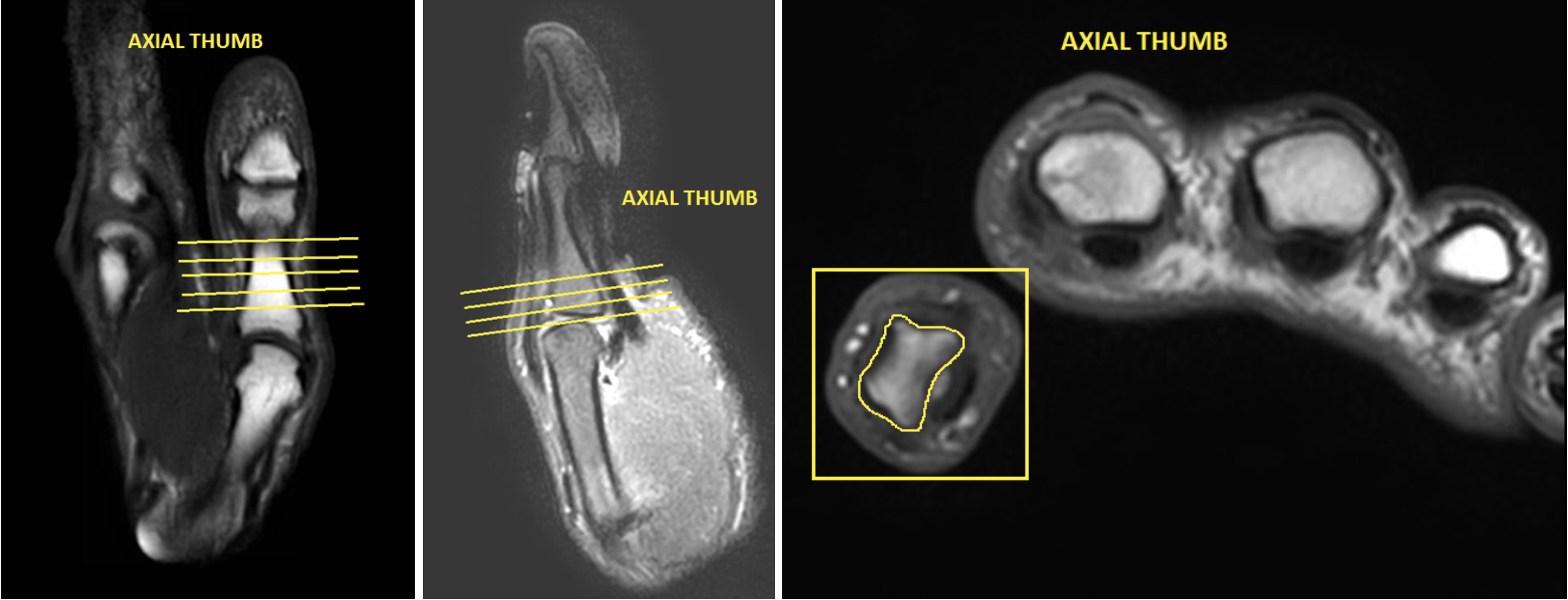
| AXIAL | T1 | TSE | 3mm | 0.5mm | None | 12cm | Cover area of interest |
| AXIAL | Mid TE (40-50) T2 Fat Sat | TSE | 3mm | 0.5mm | SPAIR | 12cm | Cover area of interest |
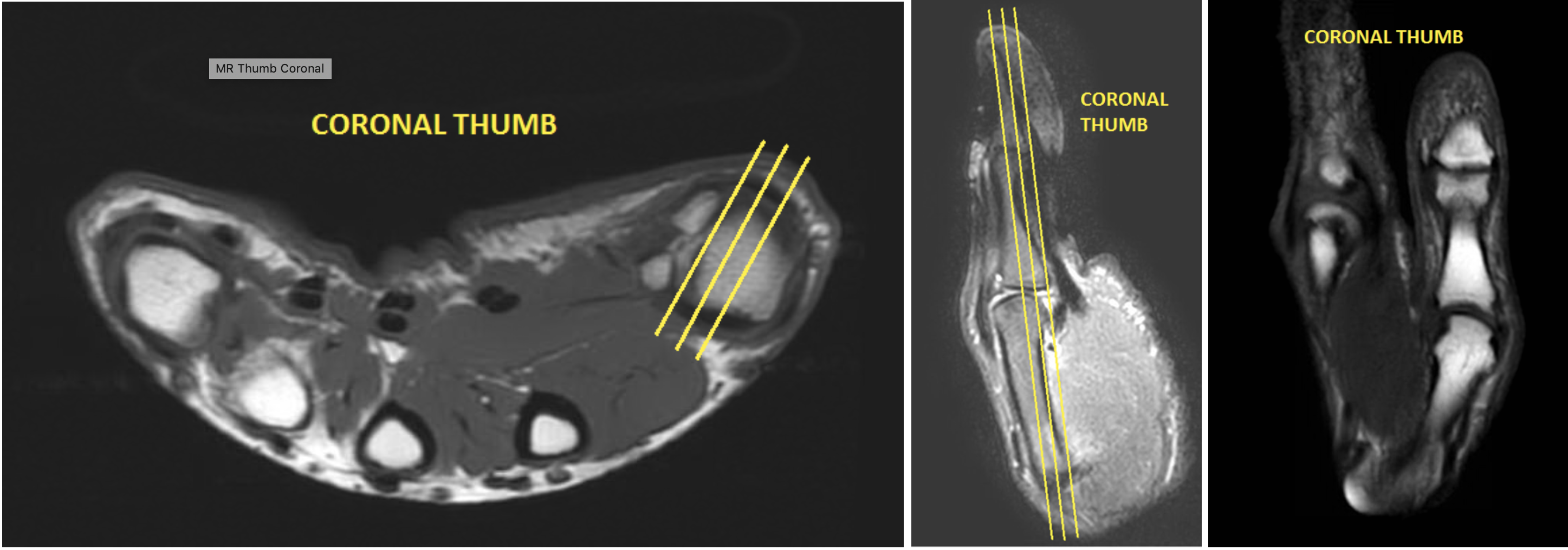
| COR | T1 | TSE | 3mm | 0.5mm | None | 12cm | Angle relative to Radio-Ulnar joint |
| COR | Mid TE (40-50) T2 Fat Sat | TSE | 3mm | 0.5mm | SPAIR | 12cm | Angle relative to Radio-Ulnar joint |
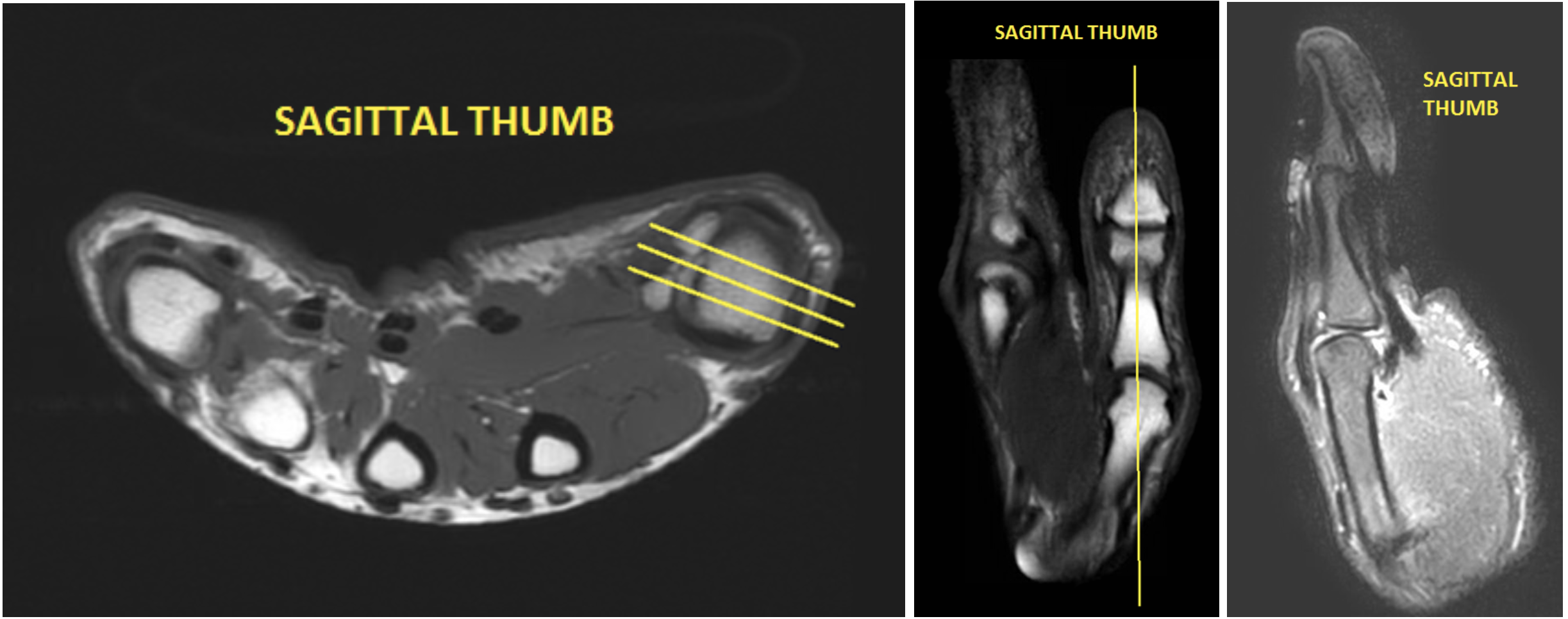
| SAG | Mid TE (40-50) T2 Fat sat | TSE | 3mm | 0.5mm | SPAIR | 12cm | Angle relative to Radio-Ulnar joint |
| SAG PRE(if giving gad for infection/osteo) | T1 | TSE | 3mm | 0.5mm | None | 12cm | Angle relative to Radio-Ulnar joint |
| POST CONTRAST | |||||||
| AXIAL | T1 Fat Sat | TSE | 3mm | 0.5mm | SPIR | 12cm | same as pre |
| COR | T1 Fat Sat | TSE | 3mm | 0.5mm | SPIR | 12cm | same as pre |
| SAG | T1 Fat Sat | TSE | 3mm | 0.5mm | SPIR | 12cm | same as pre |