MR Shoulder Arthrogram Protocol
Revised - 3/5/19
- Charge as Shoulder Arthrogram WO
- 3T Scanner Only
- Shoulder Coil
- Patients are injected in X-ray and are brought to MRI in a wheelchair by the x-ray tech.
- The screening form should be filled out.
- Images do not need to be checked by the MSK radiologist unless the tech has questions or concerns.
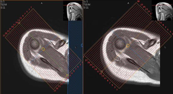
| Plane | Weighting | Mode | Slice | Gap | FAT SAT | FOV | Scan Range |
|---|---|---|---|---|---|---|---|
| AXIAL | T2 FAT SAT | TSE | 2.5mm | 0.5mm | SPAIR | 15cm | Entire Shoulder (Include A/C Joint) |
| AXIAL | T1 FAT SAT | TSE | 2.5mm | 0.5mm | SPIR | 15cm | Entire Shoulder (Include A/C Joint) |
| COR | T1 FAT SAT | TSE | 2.5mm | 0.5mm | SPIR | 15cm | Parallel to Supraspinatous |
| COR | Mid TE T2 FAT SAT | TSE | 2.5mm | 0.5mm | SPAIR | 15cm | Parallel to Supraspinatous |
| SAG | T1 FAT SAT | TSE | 2.5mm | 0.5mm | SPIR | 15cm | Perpendicular to Supraspinatous |
| SAG | T1 | TSE | 2.5mm | 0.5mm | None | 15cm | Perpendicular to Supraspinatous |
| SAG | T2 FAT SAT | TSE | 2.5mm | 0.5mm | SPAIR | 15cm | Perpendicular to Supraspinatous |
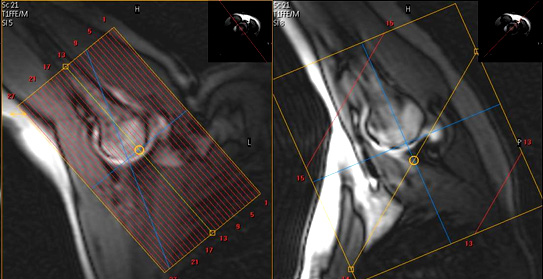
ABER localizer
Reposition patient as illustrated below for Aber. Change coil to flex coil if necessary for optimal signal. Do not perform if patient cannot tolerate position.
| Plane | Weighting | Mode | Slice | Gap | FAT SAT | FOV | Scan Range |
|---|---|---|---|---|---|---|---|
| ABER | T1 FAT SAT | TSE | 2.5mm | 0.5mm | SPIR | 15cm | ABER Oblique |